
Corneal Disease
The cornea is the transparent structure at the front of the eye, which allows images to be projected onto the retina. The cornea does not have blood vessels and depends strictly on the oxygen supply from tears and the intraocular fluid (aqueous humor).
Corneal ulcers
Corneal ulcer (ulcerative keratitis) is a condition characterized by damage to one or multiple corneal layers. Due to its delicate structure (thickness of 0.5 mm), the cornea is very sensitive to trauma and inadequate oxygenation. Since numerous bacteria normally live on the corneal surface, any damage to the corneal structure may result in rapid bacterial infection, ultimately leading to corneal destruction. Corneal ulcers may be shallow, involving only the most superficial layers of the cornea, or may be very deep, damaging nearly all of the corneal layers. Shallow ulcers are usually treated medically, while deep corneal ulcers may require surgery to rebuild the corneal integrity.
Shallow ulcers normally heal within 5–7 days. If complete ulcer healing is not present within 7–10 days, these ulcers become non-healing, or indolent, ulcers. Indolent ulcers can't heal with the use of medical therapy alone, and high-speed corneal burring or needle scraping of the ulcer surface is usually performed to stimulate healing. This procedure is usually done with the use of topical anesthetic drops or general sedative agents.
The most frequent cause of corneal ulcers is poor quality or quantity of tears, which is frequently present in dogs with seasonal or food allergies affecting the eyes, eyelids, and skin in general. Abnormal shape of the eyelids and foreign bodies are also among the causes of corneal ulcers in dogs. Symptoms of corneal ulcers usually include squinting, excessive eye redness and discharge, sensitivity to light, and corneal cloudiness (hazy or blue color).
If recognized early, most corneal ulcers heal quickly with aggressive medical therapy. Deep ulcers are almost always infected and may require hospitalization for hourly medical treatment over several days. Corneal ulcers that destroy more than 60–70% of the corneal structure usually require surgical treatment with the goal of restoring corneal integrity and preventing loss of the eye. It is very important that rubbing of an injured eye is prevented with the use of an Elizabethan protective collar — excessive rubbing on an injured eye can cause more damage to the cornea and bring more bacteria from the paws to the eye.
Corneal ulcers in cats
Feline herpesvirus infection is the most frequent cause of corneal ulcers in cats. Feline herpesvirus ulcers are usually superficial and do not respond to antibiotic therapy. If treated with topical or systemic steroids, these ulcers can get dramatically worse in a short period of time. Treatment of these ulcers is pursued with the use of anti-viral medications. Deep corneal ulcers are most frequently associated with bacterial infections and may require aggressive medical therapy. If a corneal ulcer is very deep, surgery may be needed to reinforce the corneal tissue and prevent the eye from rupturing.
Feline eosinophilic keratitis
Feline eosinophilic keratitis is an autoimmune condition that usually affects the cornea or surrounding ocular tissues in cats. This condition is characterized by the development of creamy-white deposits on the eye surface, and it usually results in eye redness and discomfort. Diagnosis is confirmed by evaluating the cells from the deposits under a microscope (cytology). Treatment is usually pursued with immunosuppressive eye drops. Since these eye drops can be very dangerous to the feline eye, all patients must be carefully monitored during treatment. In some cats, resolution of eosinophilic keratitis may be followed by reactivation of a herpes virus infection, warranting treatment with anti-viral medications.
Dermoid
A dermoid is the presence of normally-haired skin in locations where such tissue should not exist. It can be found on the cornea, limbus (the transition between the cornea and the sclera), conjunctiva, third eyelid, or eyelids. This can be a hereditary condition in some dog and cat breeds, and becomes noticeable immediately after birth or when the animal opens its eyes. The hairs act as irritants to the eye structures, causing eyelid spasms and tearing. Corneal ulcers or corneal scarring are often observed.
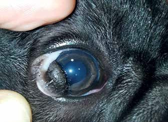
Dermoids are generally superficial and are easily removed surgically. A deeper lesion that has formed on the cornea may require surgical reconstruction using different types of graft materials to preserve the integrity of the eye globe. In general, surgical removal of a dermoid is 100% successful, and the sooner it is removed, the less the risk of more serious corneal complications. If the dermoid is removed from the eyelids, it may sometimes be necessary to perform eyelid reconstruction to preserve the anatomical structure.
Corneal dystrophy
Corneal dystrophy is a term that describes multiple different pathological conditions, which can be hereditary and progressive, leading to the appearance of minor or major localized opacities on the cornea. Corneal dystrophy is usually present in both eyes, though asymmetric presence can be observed as well. Uncontrolled allergies and other diseases or metabolic disorders — such as pancreatitis, hypothyroidism, and Cushing's disease — can worsen corneal dystrophic changes, as can systemic and topical use of steroids.
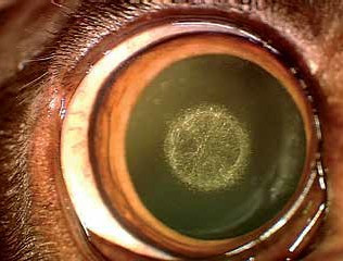
Characteristic changes on the cornea occur as a result of abnormal corneal metabolism, during which deposits of cholesterol, lipids, and calcium form. These are mostly superficial, and less frequently penetrate deeper into the corneal stroma. They appear as individual white or grayish deposits of various shapes and textures, sometimes multiple and punctate. There are no signs of eye inflammation and no corneal neovascularization (new blood vessel growth). Many dog breeds are prone to some form of corneal dystrophy, most commonly Siberian Huskies, Beagles, Shetland Sheepdogs, Cavalier King Charles Spaniels, Cocker Spaniels, Bichons, and German Shepherds. The corneal deposits do not significantly affect the quality of vision and usually do not cause eye irritation. Sometimes, brittle crystalline deposits of calcium may break off from the cornea, leaving behind a painful ulcer that may become infected. In dogs where corneal dystrophy results in recurrent ulcerations, pain, and ocular discomfort, surgical resection of the dystrophic regions is indicated.
Diagnosis is made based on examination with a slit lamp. A complete blood count, biochemistry, and hormonal status should be examined to rule out systemic diseases that could cause similar changes. Therapy usually focuses on improving the quality of the tear film and controlling allergic eye disease. In the case of complications from calcium deposition on the cornea, surgical intervention is advised.
Corneal endothelial dystrophy and degeneration
Corneal endothelial dystrophy is a progressive condition in which there is a reduction in the number of endothelial cells, as well as changes in the appearance and function of the remaining cells. Corneal endothelial cells maintain water flow within the cornea, and when this role is impaired, fluid accumulates and corneal edema occurs. Older dogs of specific breeds (Boston Terriers, Dachshunds, Chihuahuas, Miniature Poodles) are prone to this disease. Changes are noticed in both eyes, although not always of the same intensity, and initially do not significantly affect vision quality. As the disease progresses, fluid can organize and form small blisters (bullae) that, when they burst, leave very painful ulcers on the cornea. With disease progression, the eye becomes cloudy and bluish, and vision gradually deteriorates.
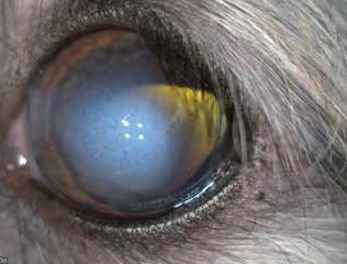
Corneal endothelial degeneration can also develop as an acquired condition, occurring after surgical procedures (phacoemulsification) or as a result of diseases that mechanically damage the corneal endothelium (glaucoma, anterior lens luxation, an intraocular tumor in contact with corneal tissue, or excessive iris cyst accumulation). With aging, the number of endothelial cells also progressively decreases, eventually reaching a point where the endothelium can no longer maintain corneal stromal dehydration, resulting in corneal edema.
In early stages, when symptoms are minimal, therapy is medical — topical ointments of hypertonic sodium chloride and anti-inflammatory drugs. If the disease has progressed, surgical therapy is advised. Various surgical procedures are used: some involve removing the superficial layer of the cornea and covering the lesion with conjunctiva to help drain the excess fluid, while others replace the damaged endothelium with healthy donor tissue.
Corneal endothelitis (deep keratitis)
Corneal endothelitis represents inflammation of the corneal endothelium, characterized by corneal edema, keratic precipitates, and a reaction in the anterior eye chamber. The cause is most commonly immunological or autoimmune in nature, and in the past often occurred after infection with the herpes virus or adenovirus CAV-1 (as well as after vaccination with a live CAV-1 virus). The patient develops a rapidly bluish-colored eye due to corneal edema, with a variable degree of ocular discomfort. Due to fluid accumulation in the cornea, blisters (bullous keratopathy), ulcerative keratitis, and secondary glaucoma can occur.
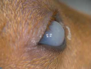
Medical history, along with clinical signs and tests for the presence of viruses, can help in making a diagnosis. Therapy includes topical and systemic administration of anti-inflammatory drugs (and steroids if indicated). The consequences of endothelitis, such as ulcers and glaucoma, must be adequately treated. In many cases, surgical intervention is required (superficial keratectomy combined with a Gundersen conjunctival graft).
Bullous keratopathy
Bullous keratopathy is the appearance of bullae on the cornea (smaller or larger blisters), which can result from corneal endothelial disease (endothelial dystrophy or endothelitis), or can develop as a result of aggressive corneal stromal inflammation and collagen degradation. If the corneal endothelium is unable to maintain normal fluid transport, excess fluid accumulates in the cornea, followed by edema and the formation of bullae. Less commonly, a bulla on the cornea can be observed in infectious keratitis (keratomalacia), where enzymes originating from bacteria, damaged corneal cells, and inflammatory cells damage the corneal structure, making it thinner, softer, and prone to bullae formation. Bullous keratopathy in cats occurs as a result of an aggressive immune response, where the immune system attacks corneal collagen, and generally occurs in cats receiving topical or systemic immunosuppressive drugs.
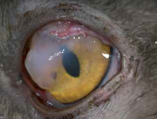
When the bullae burst, very painful ulcers can form. They can become infected, and the process can extend to the deeper layers of the cornea, leading to the formation of a descemetocele, where only Descemet's membrane prevents eye rupture. In some cases, a very aggressive course of disease results in corneal perforation and the need for emergency corneal repair surgery.
Therapy aims to stabilize enzymatic processes using serum, tetracyclines, EDTA solutions, and N-acetyl cysteine. Local anticollagenases and antibiotics are applied. Surgically, the edematous corneal tissue can be covered with conjunctiva or a third eyelid flap (feline bullous keratopathy). If the bullae formed due to endothelial dystrophy, the therapy indicated for endothelial dystrophy is applied.
More in the Atlas of Ocular Diseases
Dealing with a painful or cloudy eye?
Corneal problems can worsen quickly — please don't wait to have it evaluated.