
Stromal Ulcer
Unlike epithelial ulcers, which involve only the corneal epithelium and are generally not visible without fluorescein, stromal ulcers extend into the corneal stroma and vary in depth, shape, and size. They can therefore be seen without fluorescein staining. They are accompanied by a marked reaction of surrounding tissue, corneal edema, and later corneal neovascularization.
Stromal ulcers may be caused by infectious agents; anatomic abnormalities such as ectopic cilia, entropion, scar tissue, or eyelid neoplasia; qualitative and/or quantitative tear-film deficiency; neurologic dysfunction affecting innervation of the cornea and/or eyelids; and trauma. Regardless of the primary cause, secondary bacterial infection commonly complicates the clinical picture. A sample should therefore always be obtained for microbiologic and cytologic examination. Blepharospasm, increased tearing, and yellow or green ocular discharge are typical. Brachycephalic dogs are particularly predisposed to stromal ulcers.
Treatment
Topical antibiotics are selected according to cytologic and microbiologic findings. If an ulcer is nonprogressive and involves up to 50% of corneal thickness, medical treatment alone may be sufficient. A progressive ulcer is an ophthalmic emergency that threatens vision and may result in corneal perforation. In addition to medical treatment, the corneal defect is surgically supported with a conjunctival, amniotic, or corneal graft, or by corneoscleroconjunctival transposition.
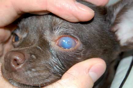
Stromal ulcer in a dog. Corneal edema is pronounced and the floor of the ulcer is slightly darker than the surrounding cornea, indicating that the cornea is very thin and at risk of perforation.
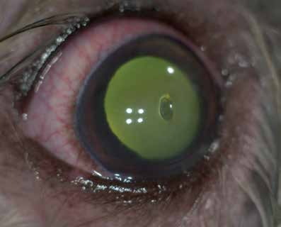
Stromal ulcer in a dog. Conjunctival hyperemia is pronounced.
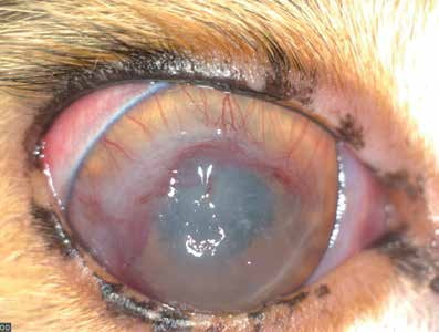
Stromal ulcer in a cat. A large stromal ulcer, anterior uveitis, and fibrin and blood within the anterior chamber are visible.
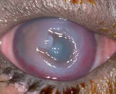
Stromal ulcer in a cat with marked corneal edema and development of deep corneal blood vessels.
Related in-depth guide
Corneal Disease
Our full guide, including treatment and surgical options
Adapted from the Atlas of Ocular Diseases in Dogs and Cats by Dr. Siniša D. Grozdanić and colleagues. © Animal Eye Consultants of Iowa. The text and clinical images on this page are original content from the authors' own clinical practice and research, and may not be reproduced, distributed, or repurposed without permission.
Think your pet may have stromal ulcer?
Early evaluation gives us the most treatment options and the best chance of preserving vision.