
Corneal Sequestrum in Cats
Corneal sequestrum is commonly seen in cats, particularly Persian and Himalayan cats. It may occur as a primary disease, but more commonly develops after chronic corneal ulceration, keratitis associated with FHV-1, or chronic irritation caused by entropion or trichiasis. It may also occur after grid keratotomy or after topical corticosteroid therapy.
A corneal sequestrum is an area of corneal degeneration containing necrotic keratocytes, altered collagen, and inflammatory cells, often with deposition of brown to black melanin. The lesion may be superficial or may extend deeply through the cornea to Descemet's membrane. Vascularization may be present. Dark sequestral material may occasionally separate from the cornea, leaving an ulcer of variable depth. Pain, blepharospasm, and ocular discharge may be present.
Treatment
Treatment is surgical, with removal of the sequestrum by keratectomy. If required, a conjunctival graft or corneoconjunctival transposition is performed. The source describes corneoconjunctival transposition as having the highest success rate for permanent resolution. Topical antibiotics and, when indicated, antiviral medications are administered.
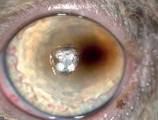
Corneal sequestrum in a cat. A large erosion and corneal pigmentation are present. Corneal blood vessels are not visible early in the disease.
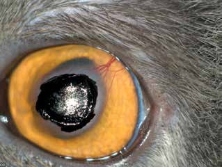
Corneal sequestrum in a cat. A network of corneal blood vessels has developed and extends toward the sequestrum.
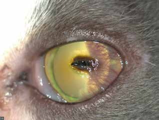
Corneal sequestrum in a cat. In this cat, the sequestrum caused an ulcer that subsequently became bacterially contaminated. A branching network of corneal blood vessels, corneal edema, and ocular discharge are present.
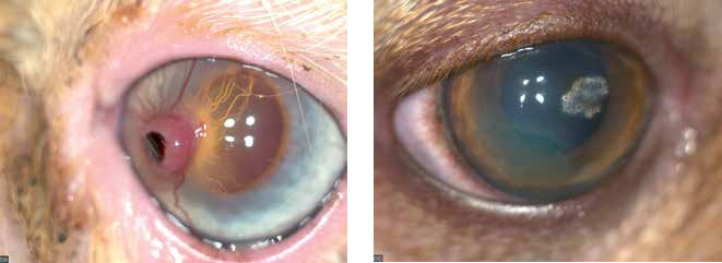
Corneal sequestrum in a cat. The sequestrum is slowly separating from the corneal tissue and will leave an ulcer after detachment.
Related in-depth guide
Corneal Disease
Our full guide, including treatment and surgical options
Adapted from the Atlas of Ocular Diseases in Dogs and Cats by Dr. Siniša D. Grozdanić and colleagues. © Animal Eye Consultants of Iowa. The text and clinical images on this page are original content from the authors' own clinical practice and research, and may not be reproduced, distributed, or repurposed without permission.
Think your pet may have corneal sequestrum in cats?
Early evaluation gives us the most treatment options and the best chance of preserving vision.