
Descemetocele
A descemetocele is a deep corneal ulcer that has destroyed the epithelium and stroma and reached Descemet's membrane. At that point, only Descemet's membrane together with the corneal endothelium prevents rupture of the globe. This condition develops through progression of a deep stromal ulcer or after trauma and is an ophthalmic emergency because of the risk of corneal perforation.
Because Descemet's membrane is a thin membrane exposed to pressure from the aqueous humor and intraocular structures without support from the damaged corneal layers, it prolapses anteriorly toward the center of the ulcerative lesion. Even small tears in Descemet's membrane may cause aqueous humor leakage, entry of microorganisms into the anterior chamber, and formation of anterior synechiae.
The patient may have pronounced discomfort and ocular pain, with blepharospasm, increased tearing, or abnormal ocular discharge. It is important to remember that many patients do not demonstrate obvious ocular pain, which may lead owners to underestimate the urgency of the condition.
Treatment
A descemetocele appears as a deep corneal ulcer whose center does not retain fluorescein stain. Because these patients are at risk of globe rupture, all manipulation must be performed carefully and surgical stabilization should be performed as soon as possible to provide secure mechanical support for the intraocular structures. Various conjunctival graft techniques may be used when the corneal lesion is smaller than 5 mm, although conjunctiva alone does not provide ideal mechanical support. Corneal grafts, whether frozen or fresh and autologous or donor-derived, synthetic grafts, or corneoscleroconjunctival transposition provide stronger support.
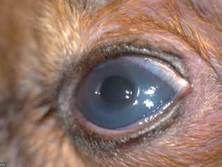
Descemetocele in a dog.
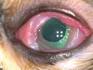
Descemetocele in a cat with pronounced conjunctival swelling and hyperemia.
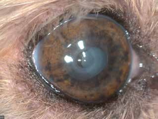
Descemetocele in a dog. Note the absence of signs of ocular irritation because the owner had been using topical corticosteroids, masking signs of inflammation.
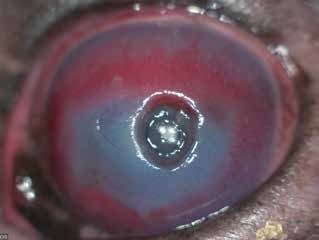
Descemetocele in a dog. The floor of the ulcer is a smooth surface, indicating that no stromal layer remains and only Descemet's membrane is visible. Deep corneal blood vessels are present.
Related in-depth guide
Corneal Disease
Our full guide, including treatment and surgical options
Adapted from the Atlas of Ocular Diseases in Dogs and Cats by Dr. Siniša D. Grozdanić and colleagues. © Animal Eye Consultants of Iowa. The text and clinical images on this page are original content from the authors' own clinical practice and research, and may not be reproduced, distributed, or repurposed without permission.
Think your pet may have descemetocele?
Early evaluation gives us the most treatment options and the best chance of preserving vision.