
Phacolytic Uveitis
Phacolytic uveitis is inflammation of the uveal tract caused by leakage of lens proteins through an intact lens capsule. It occurs in dogs with cataracts, particularly when cataract resorption begins. It frequently accompanies diabetic cataracts that develop suddenly and progress rapidly.
Clinical signs resemble chronic uveitis and include conjunctival and episcleral hyperemia, corneal edema, keratic precipitates on the endothelium, iris hyperpigmentation, aqueous flare, miosis, and decreased intraocular pressure. Synechiae, secondary glaucoma, and retinal detachment are common complications.
Treatment
Diagnosis is established by ophthalmic examination. Uveitis is treated with topical and, when necessary, systemic anti-inflammatory medications. If secondary complications have not caused permanent loss of vision, surgical lens removal by phacoemulsification is recommended because it removes the primary source of inflammation.
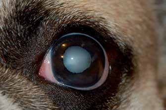
Phacolytic uveitis secondary to cataract in a young dog. Uveal ectropion is visible.
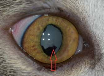
Phacolytic uveitis in a young cat. Uveal ectropion and posterior synechiae, red arrow, are visible.
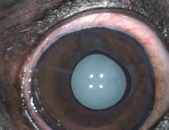
Phacolytic uveitis in a young dog.
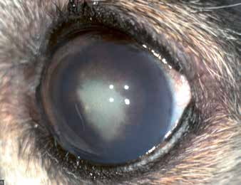
Phacolytic uveitis in a dog with posterior synechiae.
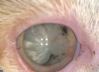
Chronic phacolytic uveitis in a cat. Posterior synechiae, pigmentation of the lens capsule, and corneal vascularization are visible.
Related in-depth guide
Cataracts
Our full guide, including treatment and surgical options
Adapted from the Atlas of Ocular Diseases in Dogs and Cats by Dr. Siniša D. Grozdanić and colleagues. © Animal Eye Consultants of Iowa. The text and clinical images on this page are original content from the authors' own clinical practice and research, and may not be reproduced, distributed, or repurposed without permission.
Think your pet may have phacolytic uveitis?
Early evaluation gives us the most treatment options and the best chance of preserving vision.