
Phacoclastic Uveitis
Phacoclastic uveitis develops when the lens capsule ruptures and the body produces a severe acute intraocular inflammatory reaction to normally sequestered lens proteins. Any penetrating trauma can damage the lens capsule. The most common cause described in the source is a cat scratch, particularly in young, inexperienced dogs.
After penetrating trauma, a localized cataract may form at the site of lens injury, or a diffuse progressive cataract may develop secondary to marked inflammation. Not every penetrating lens injury causes complications. Phacoclastic uveitis is more likely when the capsular defect is larger, the lens cortex is damaged, or secondary bacterial infection develops.
Spontaneous lens-capsule rupture may also occur. In dogs with diabetes and rapidly progressive diabetic cataracts, spontaneous equatorial rupture can be seen. Posterior spontaneous capsular rupture can also occur in nondiabetic cataracts, especially in young dogs with intumescent cataracts.
Regardless of the cause of capsular rupture, phacoclastic uveitis presents as severe uveitis with conjunctival and episcleral hyperemia, corneal edema, endothelial keratic precipitates, iris hyperpigmentation, aqueous flare, miosis, and reduced intraocular pressure. Hyphema or hypopyon may occur. Synechiae, secondary glaucoma, and retinal detachment are frequent complications.
Treatment
Diagnosis is based on the history and ophthalmic examination. A corneal wound from prior perforation may be visible. Injury to the lens capsule may be more difficult to identify, but fibrin and inflammatory material may be deposited at the capsular lesion. Topical and systemic anti-inflammatory medications are used, although the source notes that response to medical therapy is often poor. Treatment is surgical, but uveitis may persist for months after cataract surgery and reduce surgical success. If treatment is delayed, multiple complications have developed, and the eye is blind and painful, enucleation is recommended.
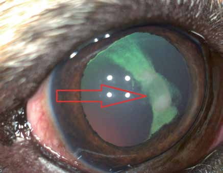
Phacoclastic uveitis in a dog after trauma to the anterior lens capsule. The arrow indicates a circular defect. Posterior synechiae, fibrin, and cortical lens material are present in the anterior chamber.
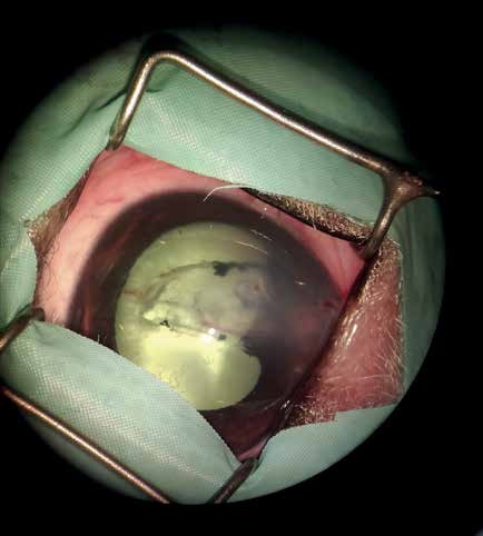
Phacoclastic uveitis in a dog caused by traumatic cataract after laceration of the anterior lens capsule by a cat claw.
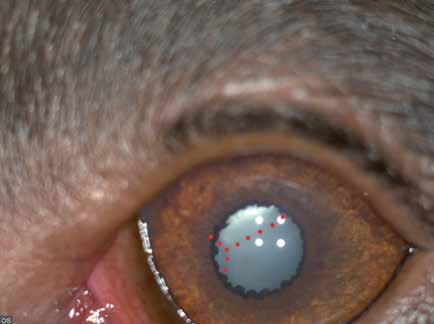
Phacoclastic uveitis in a young dog with an intumescent cataract secondary to posterior lens-capsule rupture. Red dots highlight displaced Y-shaped lens sutures; when these are not centrally located, they may indicate capsular rupture.
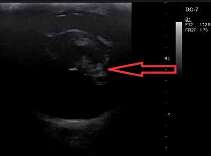
Ultrasound examination of phacoclastic uveitis in the dog shown in the previous figure. The arrow indicates the lens defect and cortical material extending into the vitreous.
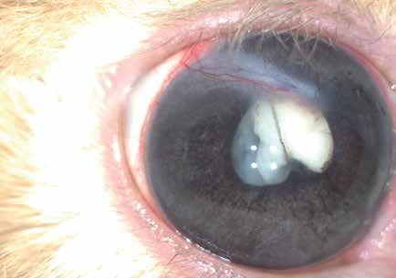
Phacoclastic uveitis following traumatic cataract caused by a cat scratch. Whitish scar tissue is visible at the site of the corneal laceration.
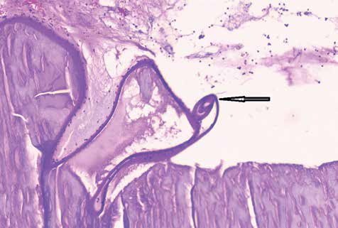
Histopathologic preparation from a dog with phacoclastic uveitis. Severe uveitis led to secondary glaucoma, blindness, and loss of the eye. No corneal wound, lens-capsule injury, or traumatic cataract was visible before enucleation. Histopathology revealed numerous neutrophils within the lens, raising suspicion of capsular rupture. After extensive sectioning, a small traumatic defect was identified at the lens equator, including discontinuity and a spiraled segment of capsule. In this case, a cat claw had passed through the sclera and produced a small equatorial lens injury that was not clinically visible and did not produce an obvious traumatic cataract, yet it was sufficient to permit lens-protein leakage and cause phacoclastic uveitis.
Related in-depth guide
Cataracts
Our full guide, including treatment and surgical options
Adapted from the Atlas of Ocular Diseases in Dogs and Cats by Dr. Siniša D. Grozdanić and colleagues. © Animal Eye Consultants of Iowa. The text and clinical images on this page are original content from the authors' own clinical practice and research, and may not be reproduced, distributed, or repurposed without permission.
Think your pet may have phacoclastic uveitis?
Early evaluation gives us the most treatment options and the best chance of preserving vision.