
Limbal Melanoma
Melanocytic tumors are relatively common at the canine limbus. Most are melanocytomas, benign tumors of melanocytes; however, the term “limbal melanoma” is commonly used in the literature regardless of whether the tumor is benign or malignant. Although benign, a limbal melanocytoma can be locally invasive and extend into the cornea or intraocular structures. It is often observed in dogs with darker pigmentation, such as German Shepherd Dogs.
A limbal melanocytoma is usually well circumscribed, elevated, and darkly pigmented, although it may rarely be nonpigmented (amelanotic). It most commonly occurs in two age groups. When it develops in dogs two to four years of age, the source describes it as rapidly progressive and highly invasive. When it occurs in older dogs eight to eleven years of age, it is generally nonprogressive and often found incidentally during ophthalmic examination. The source suggests a partially hereditary component in some breeds, including Labrador and Golden Retrievers. It occurs rarely in cats.
Treatment
Limbal melanocytoma must be differentiated diagnostically from primary uveal melanoma that has extended to the limbus. A detailed ophthalmic examination, gonioscopy, ultrasonography, and ultimately biopsy are important. Definitive diagnosis is established by histopathologic examination after excision. Although the tumor is generally benign, the source recommends a comprehensive systemic evaluation including complete blood count, serum biochemistry, and radiography. Treatment consists of surgical removal, with placement of a conjunctival or corneoscleral graft. Recurrence is very uncommon after successful surgery.
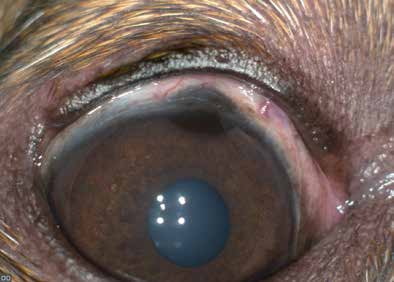
Melanocytoma (melanoma) of the canine limbus.
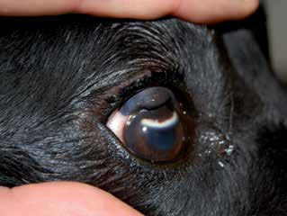
Large melanocytoma (melanoma) of the canine limbus. The whitish arcuate lesion beneath the melanoma is an area of corneal degeneration.
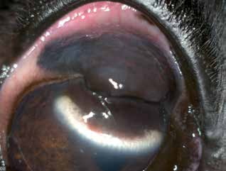
Melanocytoma (melanoma) of the limbus in the same dog as in the previous figure, after retraction of the upper eyelid.
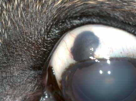
Melanocytoma (melanoma) of the canine limbus.
Related in-depth guide
Corneal Disease
Our full guide, including treatment and surgical options
Adapted from the Atlas of Ocular Diseases in Dogs and Cats by Dr. Siniša D. Grozdanić and colleagues. © Animal Eye Consultants of Iowa. The text and clinical images on this page are original content from the authors' own clinical practice and research, and may not be reproduced, distributed, or repurposed without permission.
Think your pet may have limbal melanoma?
Early evaluation gives us the most treatment options and the best chance of preserving vision.