
Keratomalacia (Acute Necrotizing Ulcer)
Keratomalacia is a complication of a corneal ulcer in which acute degradation of the corneal stroma occurs. During normal corneal wound healing, a certain amount of protease and collagenase activity is released to remove damaged tissue. In keratomalacia, large quantities of proteases and collagenases are released by corneal epithelial cells, fibroblasts, polymorphonuclear leukocytes, and some microorganisms.
Accumulation of these enzymes causes progressive and acute breakdown of the stroma, which develops a gelatinous appearance and is often displaced anteriorly, creating a bullous formation. Pronounced neovascularization and varying degrees of corneal edema may be present, and corneal perforation is a common complication. Patients show blepharospasm and mucoid ocular discharge. The disease is diagnosed more frequently in Shih Tzus, Pugs, Lhasa Apsos, Boston Terriers, and other brachycephalic dog breeds.
Treatment
Treatment consists of frequent topical administration of antibiotics and serum, EDTA solution, and N-acetylcysteine to stabilize proteolytic activity. Doxycycline is used systemically. In exceptionally advanced cases, altered gelatinous tissue must be removed and a conjunctival graft placed to prevent globe rupture. The source describes corneal collagen cross-linking (CXL) as producing very good results in stabilizing the process.
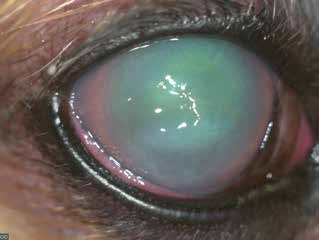
Keratomalacia in a dog. Marked corneal edema and a gelatinous appearance of the tissue are visible.
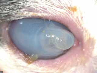
Keratomalacia in a cat with corneal perforation. A bullous formation is visible because the cornea has lost its structural strength.
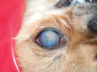
Keratomalacia in a dog with corneal perforation. The large white, button-like area represents accumulation of fibrin and necrotic corneal stroma. Marked corneal edema and blood in the anterior chamber are visible.
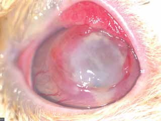
Keratomalacia in a cat without corneal perforation. The cornea has a gelatinous appearance, with superficial and deep corneal blood vessels and edematous, hyperemic conjunctiva. The condition resulted from a chronic ulcer that subsequently became contaminated.
Related in-depth guide
Corneal Disease
Our full guide, including treatment and surgical options
Adapted from the Atlas of Ocular Diseases in Dogs and Cats by Dr. Siniša D. Grozdanić and colleagues. © Animal Eye Consultants of Iowa. The text and clinical images on this page are original content from the authors' own clinical practice and research, and may not be reproduced, distributed, or repurposed without permission.
Think your pet may have keratomalacia (acute necrotizing ulcer)?
Early evaluation gives us the most treatment options and the best chance of preserving vision.