
Fungal Keratitis
Fungal keratitis is diagnosed relatively rarely in dogs and cats. Aspergillus spp. are described as the most common cause in the source. The condition generally develops after epithelial trauma or immunosuppression, particularly after prolonged topical treatment with antibiotics and/or corticosteroids. Lesions may occasionally be ulcerative, but irregular whitish-yellow or grayish corneal deposits are almost always visible. Keratitis is progressive and is often complicated by bacterial infection, with secondary keratomalacia and corneal perforation.
Treatment
Diagnosis can be established by cytologic or histologic examination of a representative corneal sample, as well as by isolation of the microorganism using culture media suitable for fungal growth. Treatment consists of topical antifungal medications. Because bacterial coinfection is common, antibiotics and often anti-inflammatory medications are also required. In some cases, successful treatment requires surgical removal of affected corneal tissue and placement of a conjunctival graft.
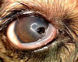
Fungal keratitis in a dog. A deep corneal ulcer with dark fungal deposits within the ulcer is visible.
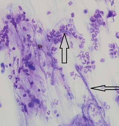
Cytologic preparation from the dog shown in the previous figure. Hyphae of Paecilomyces sp. are indicated by black arrows and are surrounded by inflammatory cells.
Related in-depth guide
Corneal Disease
Our full guide, including treatment and surgical options
Adapted from the Atlas of Ocular Diseases in Dogs and Cats by Dr. Siniša D. Grozdanić and colleagues. © Animal Eye Consultants of Iowa. The text and clinical images on this page are original content from the authors' own clinical practice and research, and may not be reproduced, distributed, or repurposed without permission.
Think your pet may have fungal keratitis?
Early evaluation gives us the most treatment options and the best chance of preserving vision.