
Epithelial Inclusion Cysts
Epithelial cysts can occasionally form at the site of a healed corneal injury or after corneal surgery as a result of transplantation of epithelial cells into the corneal stroma. The cysts slowly enlarge as proteinaceous material and debris accumulate within them. They may occasionally occur as a congenital anomaly. They appear as localized, round, smooth, white-yellow, pink, or brown structures elevated above the cornea at the site of a previous injury. Moderate neovascularization is present, and the lesion itself generally causes little discomfort.
Treatment
Treatment is required if a cyst enlarges or irritates the cornea. It is removed surgically by superficial keratectomy, with placement of a conjunctival graft. Tissue should be submitted for histopathologic examination to confirm the diagnosis. Topical antibiotics and anti-inflammatory medications are used postoperatively. In some cases, it is sufficient to excise only the portion of the cyst protruding above the corneal surface with a corneal knife and then treat the resulting lesion as an ulcer.
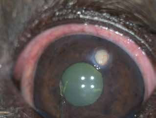
Epithelial inclusion cyst on the cornea of a dog.
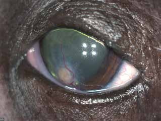
Epithelial inclusion cyst on the cornea of a dog that developed as a complication of previous corneoscleral transposition surgery.
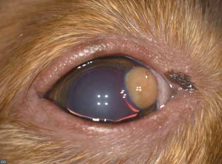
Epithelial inclusion cyst on the cornea of a dog. After removal of the cyst, a descemetocele was discovered, which is unusual for this condition.
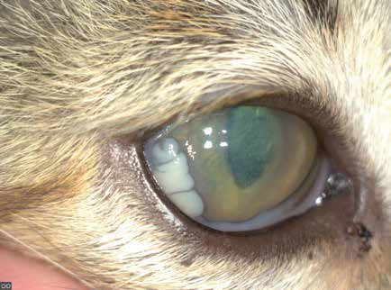
Multiple epithelial inclusion cysts on the cornea of a cat after herpesvirus infection.
Related in-depth guide
Corneal Disease
Our full guide, including treatment and surgical options
Adapted from the Atlas of Ocular Diseases in Dogs and Cats by Dr. Siniša D. Grozdanić and colleagues. © Animal Eye Consultants of Iowa. The text and clinical images on this page are original content from the authors' own clinical practice and research, and may not be reproduced, distributed, or repurposed without permission.
Think your pet may have epithelial inclusion cysts?
Early evaluation gives us the most treatment options and the best chance of preserving vision.