
Corneoscleral (Limbal) Hemangioma/Hemangiosarcoma
Tumors of vascular endothelial origin, including hemangioma and hemangiosarcoma, may involve corneoscleral as well as conjunctival tissue. They appear as elevated, red, smooth, irregularly shaped masses, most commonly located near the limbus. Exposure to ultraviolet radiation is considered an important factor in their development.
Hemangioma is benign, whereas hemangiosarcoma is malignant. Hemangioma is well demarcated from surrounding tissue and generally causes little difficulty, while hemangiosarcoma is locally invasive and can destroy adjacent tissue. Metastases are uncommon. These tumors are reported somewhat more frequently in Australian Shepherds and Collies and occur rarely in cats.
Treatment
Definitive information about tumor type and malignancy is obtained by histopathologic examination. Surgical excision and cryotherapy may be required. Enucleation may occasionally be necessary if hemangiosarcoma is very invasive. The source describes favorable results following perilesional administration of anti-VEGF therapy (Avastin).
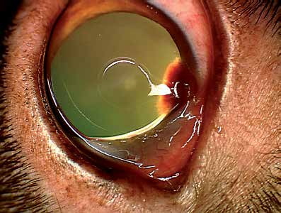
Limbal hemangioma/hemangiosarcoma in a dog.
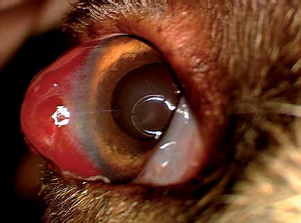
Hemangioma/hemangiosarcoma at the canine limbus.
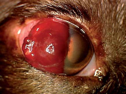
Corneoscleral hemangioma/hemangiosarcoma in a dog.
Related in-depth guide
Corneal Disease
Our full guide, including treatment and surgical options
Adapted from the Atlas of Ocular Diseases in Dogs and Cats by Dr. Siniša D. Grozdanić and colleagues. © Animal Eye Consultants of Iowa. The text and clinical images on this page are original content from the authors' own clinical practice and research, and may not be reproduced, distributed, or repurposed without permission.
Think your pet may have corneoscleral (limbal) hemangioma/hemangiosarcoma?
Early evaluation gives us the most treatment options and the best chance of preserving vision.