
Corneoscleral Lacerations
A traumatic corneoscleral laceration is a relatively common ophthalmic emergency.
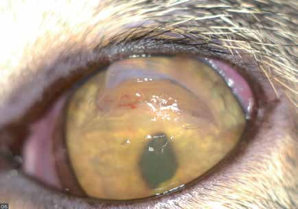
Corneal laceration in a cat. Whitish fibrin deposits are visible at the edge of the laceration, together with corneal edema and miosis.
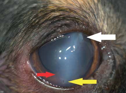
Corneal laceration caused by a cat scratch. The white arrow indicates the entry wound and the yellow arrow the exit wound made by the cat's claw. The red arrow indicates anterior synechiae and altered iris anatomy because the iris was also in the path of the claw. Uveal entropion is also visible.
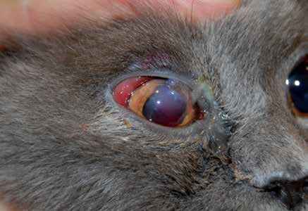
Corneal laceration in a cat. A corneal defect and iris prolapse, hyphema, deformation of the pupil, swollen hyperemic conjunctiva, and purulent discharge are visible.
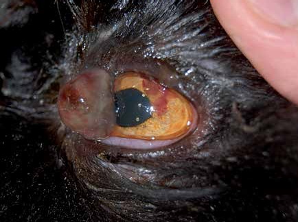
Ocular laceration in a cat. Iris prolapse, blood and fibrin in the anterior chamber, and deformation of the pupil are present.
The most common cause of these injuries is a cat scratch. A laceration may be superficial or may penetrate all corneal layers, resulting in leakage of aqueous humor and collapse of the anterior chamber. Lacerations extending through no more than one third of the corneal thickness and located in the peripheral cornea have a better prognosis. Deeper lacerations that perforate the cornea may cause aqueous humor leakage, anterior synechiae, or iris prolapse.
If the lens capsule is injured, lens proteins leak from the lens and provoke a severe autoimmune reaction and subsequent uveitis because the body recognizes them as foreign material. Even a very small lens injury can cause complications because a localized cataract may develop and eventually involve the entire lens. Hyphema and infection of the entire eye following penetration of microorganisms are also possible. The condition is very painful and causes marked tearing and blepharospasm. For these reasons, all corneal lacerations should be considered serious and a detailed ophthalmic examination should be performed immediately to determine the extent of injury.
Treatment
Diagnosis is readily made from the history, clinical presentation, and ophthalmic examination. Small superficial lacerations may heal with medical therapy, whereas more serious and deeper lacerations require surgical repair as soon as possible. Topical and systemic antibiotics and anti-inflammatory medications are administered. If the lens has been injured and a traumatic cataract has developed, phacoemulsification is performed with implantation of an artificial intraocular lens.
Related in-depth guide
Corneal Disease
Our full guide, including treatment and surgical options
Adapted from the Atlas of Ocular Diseases in Dogs and Cats by Dr. Siniša D. Grozdanić and colleagues. © Animal Eye Consultants of Iowa. The text and clinical images on this page are original content from the authors' own clinical practice and research, and may not be reproduced, distributed, or repurposed without permission.
Think your pet may have corneoscleral lacerations?
Early evaluation gives us the most treatment options and the best chance of preserving vision.