
Corneal Perforation
Corneal perforation is an ophthalmic emergency that develops because of traumatic injury, progression of a corneal ulcer, or rupture of a descemetocele. Following perforation, the patient has severe pain manifested by blepharospasm, ocular discharge, and collapse of the anterior chamber. Mucus, fibrin, and blood accumulate at the site of the wound.
Treatment
A patient with a perforated cornea should undergo surgical treatment as soon as possible to prevent complications such as iris prolapse and retinal detachment and to preserve vision and/or the eye. The defect must be repaired and lost aqueous humor replaced. Different surgical procedures are recommended depending on the type of defect. After repair using a conjunctival or corneal graft or corneoscleroconjunctival transposition, topical and systemic antibiotic therapy is administered together with nonsteroidal anti-inflammatory medication.
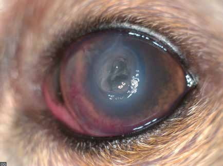
Corneal perforation following a cat scratch, with formation of a fibrin plug. Corneal edema and vascularization are present.
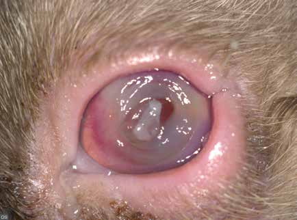
Corneal perforation in a cat. This developed after separation of a sequestrum from the corneal tissue, producing a deep stromal ulcer and subsequently corneal perforation. A fibrin plug, corneal edema, and vascularization are visible.
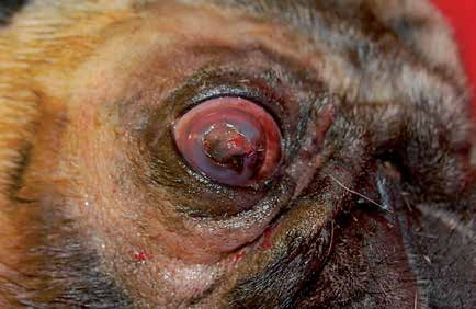
Corneal perforation with iris prolapse secondary to a chronic ulcer that the owner had treated with corticosteroids.
Related in-depth guide
Corneal Disease
Our full guide, including treatment and surgical options
Adapted from the Atlas of Ocular Diseases in Dogs and Cats by Dr. Siniša D. Grozdanić and colleagues. © Animal Eye Consultants of Iowa. The text and clinical images on this page are original content from the authors' own clinical practice and research, and may not be reproduced, distributed, or repurposed without permission.
Think your pet may have corneal perforation?
Early evaluation gives us the most treatment options and the best chance of preserving vision.