
Corneal Foreign Bodies
Whenever a painful eye with corneal injury is examined, the possibility of a foreign body should be considered. The most common foreign bodies are plant material and are usually embedded in the cornea, sclera, conjunctival fornix behind the eyelids, or behind the third eyelid. Pronounced blepharospasm, chemosis, conjunctival hyperemia, and ocular discharge are present.
Treatment
The foreign body must be carefully removed from the cornea, followed by topical antibiotic and nonsteroidal anti-inflammatory therapy.
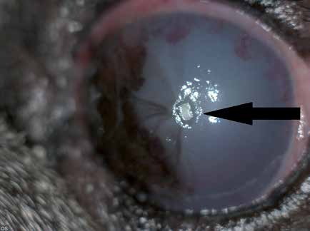
Rectangular whitish foreign body in the cornea of a dog, indicated by the arrow, that caused an ulcer. Corneal blood vessels and pigment deposition were present before the foreign body; the dog is a Pug.
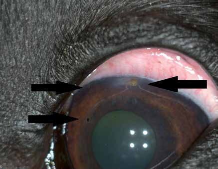
Three punctate plant-derived foreign bodies in the cornea of a dog, indicated by arrows.
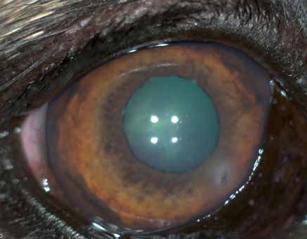
Plant thorn that perforated the cornea.
Related in-depth guide
Corneal Disease
Our full guide, including treatment and surgical options
Adapted from the Atlas of Ocular Diseases in Dogs and Cats by Dr. Siniša D. Grozdanić and colleagues. © Animal Eye Consultants of Iowa. The text and clinical images on this page are original content from the authors' own clinical practice and research, and may not be reproduced, distributed, or repurposed without permission.
Think your pet may have corneal foreign bodies?
Early evaluation gives us the most treatment options and the best chance of preserving vision.