
Corneal Endothelial Dystrophy and Degeneration
Corneal endothelial dystrophy is a progressive pathologic condition characterized by a reduction in the number of endothelial cells together with changes in corneal endothelial cell morphology and function. Corneal endothelial cells regulate water movement within the cornea. When this function is impaired by pathologic changes, fluid accumulates and corneal edema develops.
Older dogs of certain breeds are predisposed, including Boston Terriers, Dachshunds, Chihuahuas, and Miniature Poodles. Changes are present in both eyes but are not equal in severity, and early in the disease they do not substantially affect visual quality.
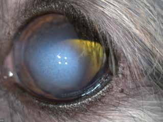
Corneal endothelial degeneration in a dog secondary to anterior lens luxation. Central corneal opacity caused by fluid accumulation is visible.
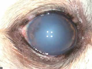
Corneal endothelial dystrophy. Most of the cornea is edematous and cloudy.
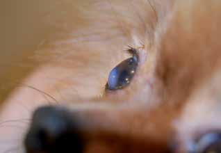
Corneal endothelial dystrophy. Localized corneal swelling is clearly visible in this dog.
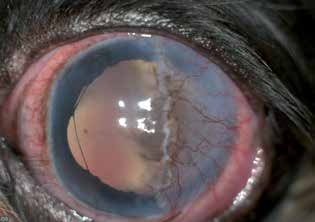
Appearance of the cornea after surgery. The purpose of this procedure is to reduce corneal thickness and bring vascularized conjunctiva to the area of corneal edema, where it can facilitate removal of fluid.
As the disease progresses, accumulated fluid may organize into small blisters, or bullae. When these rupture, they leave very painful corneal ulcers. With progression, the eye becomes cloudy and bluish and vision gradually deteriorates. Neovascularization is not visible.
Corneal endothelial degeneration is acquired and can occur after surgical procedures such as phacoemulsification, during which endothelial damage may occur, or secondary to anterior synechiae or contact between the corneal endothelium and a luxated lens or uveal mass. One or both eyes may be affected. In addition, the number of endothelial cells progressively decreases as a dog ages until a critical point is reached at which the endothelium can no longer maintain stromal dehydration, resulting in corneal edema.
Treatment
In the early stage, when clinical problems are minimal, treatment is medical and consists of topical hypertonic sodium-chloride ointments and anti-inflammatory medications. In advanced disease, treatment is surgical. Numerous surgical procedures may be indicated. Some involve removal of a superficial corneal layer and covering the lesion with conjunctiva to facilitate drainage of excess accumulated fluid; others are based on replacement of damaged endothelium with healthy donor tissue.
Related in-depth guide
Corneal Disease
Our full guide, including treatment and surgical options
Adapted from the Atlas of Ocular Diseases in Dogs and Cats by Dr. Siniša D. Grozdanić and colleagues. © Animal Eye Consultants of Iowa. The text and clinical images on this page are original content from the authors' own clinical practice and research, and may not be reproduced, distributed, or repurposed without permission.
Think your pet may have corneal endothelial dystrophy and degeneration?
Early evaluation gives us the most treatment options and the best chance of preserving vision.