
Corneal Dystrophy
Corneal dystrophy is a term describing several inherited, progressive disorders that produce smaller or larger localized corneal opacities. Corneal dystrophy is bilateral and is generally not caused by other diseases or metabolic disorders. Characteristic corneal changes result from abnormal corneal metabolism, with formation of cholesterol, lipid, and calcium deposits. These deposits are usually superficial and only rarely extend more deeply into the corneal stroma.
The lesions appear as single whitish or grayish deposits of various shapes and textures. They may also be multiple and punctate. Signs of ocular inflammation are absent and corneal neovascularization does not occur. Many dog breeds are predisposed to some form of corneal dystrophy; breeds described most commonly in the source include Siberian Huskies, Beagles, Shetland Sheepdogs, Cavalier King Charles Spaniels, Cocker Spaniels, Bichons, and German Shepherd Dogs.
Corneal deposits generally do not significantly affect vision and usually do not cause ocular irritation. Occasionally, when calcium accumulates in the corneal epithelium or stroma, brittle crystalline deposits may detach from the cornea and leave a painful ulcer that can become infected.
Treatment
Diagnosis is based on slit-lamp examination. A complete blood count, serum biochemistry profile, and hormonal status should be evaluated to exclude systemic diseases that can produce similar changes. Treatment is not necessary in mild cases. Surgical intervention is recommended when complications develop secondary to calcium deposition on the cornea.
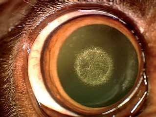
Central corneal dystrophy.
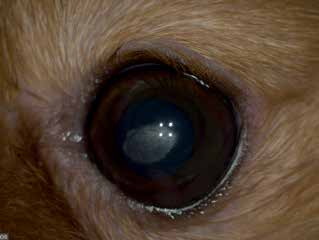
Typical position and appearance of corneal dystrophy.
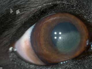
A larger area of corneal dystrophy than in the previous figures, but still without signs of ocular irritation or inflammation.
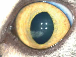
Corneal dystrophy in a cat.
Related in-depth guide
Corneal Disease
Our full guide, including treatment and surgical options
Adapted from the Atlas of Ocular Diseases in Dogs and Cats by Dr. Siniša D. Grozdanić and colleagues. © Animal Eye Consultants of Iowa. The text and clinical images on this page are original content from the authors' own clinical practice and research, and may not be reproduced, distributed, or repurposed without permission.
Think your pet may have corneal dystrophy?
Early evaluation gives us the most treatment options and the best chance of preserving vision.