
Corneal Degeneration
Corneal degeneration is a pathologic change in the cornea that develops secondary to various ocular or systemic disorders, particularly allergic diseases of the conjunctiva and eyelids, and most commonly secondary to metabolic disease such as hyperlipidemia, hypercalcemia, hypervitaminosis D, hypothyroidism, hyperadrenocorticism, and diabetes mellitus. It can also accompany diseases of the pancreas, liver, or kidneys.
Ocular diseases such as uveitis, dry eye disease, and corneal scarring can lead to degenerative corneal changes, which most commonly consist of calcium deposits. Degenerative changes are deposits of lipid or calcium and may affect one or both eyes. Neovascularization, consisting of a network of newly formed blood vessels, is usually present. Lipid deposits may occur in any corneal layer, whereas calcium deposits are generally superficial. The deposits are irregular, asymmetric, and white to gray-white in color.
Deposits may spontaneously separate from the cornea and leave painful ulcers. The dog may experience discomfort and pain, squint frequently or keep the eye closed, and show marked tearing. Mucoid ocular discharge may occasionally occur.
Treatment
A complete blood count, serum biochemistry profile, and assessment of hormonal status are used to identify the primary disorder that caused the corneal degeneration. Corneal deposits can be removed surgically. Topical EDTA solution and antibiotic ointment are used. Treatment of allergies is an important component of therapy. In addition to specific treatment for a diagnosed metabolic disorder, the diet should be adjusted to the patient's needs and condition.
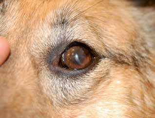
Corneal degeneration that became visible one year after corneal surgery.
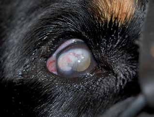
Corneal degeneration secondary to scleritis. Neovascularization is clearly visible over the white surface of the degenerative lesion.
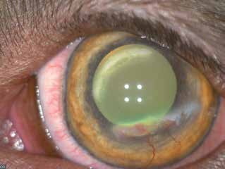
Corneal degeneration appearing as two crescent-shaped whitish lesions. Neovascularization is clearly visible on one of the lesions.
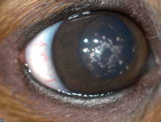
Corneal degeneration that developed after autoimmune punctate keratitis in a Dachshund.
Related in-depth guide
Corneal Disease
Our full guide, including treatment and surgical options
Adapted from the Atlas of Ocular Diseases in Dogs and Cats by Dr. Siniša D. Grozdanić and colleagues. © Animal Eye Consultants of Iowa. The text and clinical images on this page are original content from the authors' own clinical practice and research, and may not be reproduced, distributed, or repurposed without permission.
Think your pet may have corneal degeneration?
Early evaluation gives us the most treatment options and the best chance of preserving vision.