
Corneal Abscess
A corneal abscess develops when bacteria or fungi penetrate the cornea and cause infection in deeper corneal layers while the surface epithelium remains relatively intact. A yellowish corneal lesion of variable size is visible and most commonly produces a marked vascular response. Fluorescein staining is negative. The patient has pronounced blepharospasm and ocular discharge.
Treatment
Systemic and topical antibiotics, ideally selected according to culture and antimicrobial susceptibility results, are administered together with anti-inflammatory medications. If the abscess does not respond to treatment, it is removed surgically and a corneoconjunctival graft is placed.
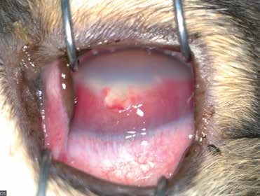
Corneal abscess in a dog. The abscess is centrally located and whitish-yellow. Pronounced corneal edema and vascularization are present, together with ocular discharge.
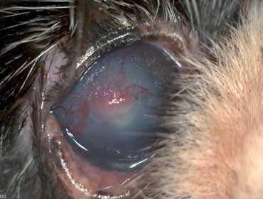
Centrally located corneal abscess in a dog, with corneal edema and vascularization.
Related in-depth guide
Corneal Disease
Our full guide, including treatment and surgical options
Adapted from the Atlas of Ocular Diseases in Dogs and Cats by Dr. Siniša D. Grozdanić and colleagues. © Animal Eye Consultants of Iowa. The text and clinical images on this page are original content from the authors' own clinical practice and research, and may not be reproduced, distributed, or repurposed without permission.
Think your pet may have corneal abscess?
Early evaluation gives us the most treatment options and the best chance of preserving vision.