
Zygomatic Gland Mucocele/Adenitis
A zygomatic gland mucocele develops because of damage to the gland or its excretory duct, leading to accumulation of saliva within a cystic structure. The process is generally unilateral, chronic, and most commonly nonpainful. Exophthalmos with protrusion of the third eyelid may be observed.
Zygomatic gland adenitis (sialoadenitis) is an acute, painful process in which a cystic structure does not form within the gland, and multiple secretory glands are often affected. The process is most commonly autoimmune in nature.
Treatment
Diagnosis is established by ultrasonography, CT, or MRI. Three to four weeks of treatment with nonsteroidal anti-inflammatory drugs may lead to resolution of swelling and may resolve the problem. Surgical resection of the cyst is also an option. Polidocanol may be injected into the cyst to induce regression, although it can occasionally cause a severe inflammatory reaction.
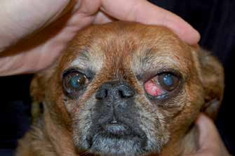
Unilateral zygomatic gland mucocele. Exophthalmos and protrusion of the third eyelid are present.
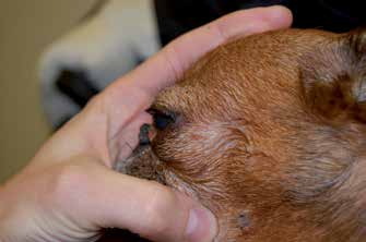
Zygomatic gland mucocele, same dog, lateral view.
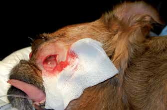
Zygomatic gland mucocele, same dog, during surgical drainage of mucoid contents.
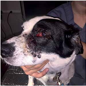
Adenitis of secretory glands. This dog has unilateral swelling on the left side of the face involving the third eyelid gland as well as the submandibular, zygomatic, and parotid glands.
Adapted from the Atlas of Ocular Diseases in Dogs and Cats by Dr. Siniša D. Grozdanić and colleagues. © Animal Eye Consultants of Iowa. The text and clinical images on this page are original content from the authors' own clinical practice and research, and may not be reproduced, distributed, or repurposed without permission.
Think your pet may have zygomatic gland mucocele/adenitis?
Early evaluation gives us the most treatment options and the best chance of preserving vision.