
Vitreous Degeneration
The vitreous is a transparent hydrogel that occupies approximately 80% of the canine globe and provides structural support to the lens and neuroretina. It consists predominantly of water, approximately 99%, while the remaining component includes fibers, hyalocytes, hyaluronic acid, and other glycosaminoglycans.
With aging, the gel structure disintegrates and becomes more liquid, while firmer fibrous particles or strands may become visible within the vitreous, a process termed vitreous syneresis. Primary vitreous degeneration is described in Griffons, Chihuahuas, Chinese Crested Dogs, Papillons, Shih Tzus, and Whippets. Intraocular inflammation, uveitis or cataract, lens luxation, and retinal atrophy can cause secondary vitreous degeneration. Liquefaction of the vitreous may allow separation from the neuroretina and can result in retinal tears and rhegmatogenous retinal detachment.
Small vitreous particles may also result from accumulation of calcium and phosphate crystals, termed asteroid hyalosis, or cholesterol crystals, termed synchysis scintillans.
Treatment
Diagnosis is established by ophthalmic examination and ultrasonography. Treatment is usually unnecessary unless retinal detachment occurs, in which case vitreoretinal surgery is required. In dogs with cataract and pronounced vitreous degeneration, phacoemulsification carries a higher risk of complications.
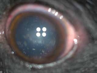
Vitreous degeneration in a dog: asteroid hyalosis.
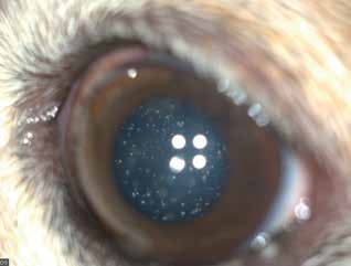
Vitreous degeneration in a dog. Numerous whitish dots within the vitreous become visible when the pupil is dilated and illumination is adequate.
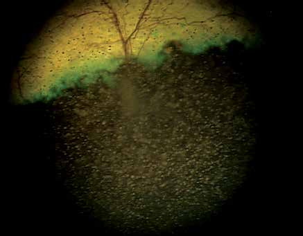
Vitreous degeneration in a dog: appearance of asteroid hyalosis on indirect ophthalmoscopy.
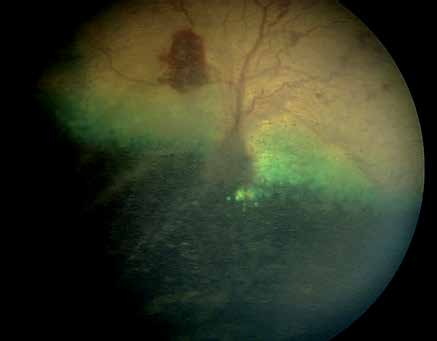
Vitreous degeneration in a dog, fundus appearance. An area of subretinal hemorrhage and retinal detachment secondary to vitreous degeneration are visible.
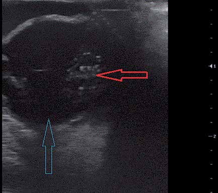
Ultrasound image of vitreous degeneration in a dog. The red arrow indicates a hyperechoic portion of the vitreous. The blue arrow indicates vitreous with normal echogenicity.
Adapted from the Atlas of Ocular Diseases in Dogs and Cats by Dr. Siniša D. Grozdanić and colleagues. © Animal Eye Consultants of Iowa. The text and clinical images on this page are original content from the authors' own clinical practice and research, and may not be reproduced, distributed, or repurposed without permission.
Think your pet may have vitreous degeneration?
Early evaluation gives us the most treatment options and the best chance of preserving vision.