
Variations in Optic Nerve Myelination
The optic nerve head is readily visible during fundic examination in dogs and cats. In cats, the optic nerve normally loses its myelin as it enters the eye, so the disc is usually consistently round, gray or white, and smaller in diameter than in dogs; variations in myelination are uncommon.
Dogs show marked variation in the amount of myelin around the optic nerve head, resulting in differences in shape and size. The canine optic nerve head may be round, oval, triangular, or polygonal. When myelination is pronounced, radiating whitish formations extend from the optic nerve head into the surrounding fundus, particularly along retinal vessels. These physiologic variations must be distinguished from pathologic changes such as optic neuritis or papilledema.
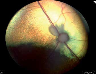
Feline fundus. Normal variation in optic disc myelination, with myelin radiating from the optic nerve head toward the tapetal region.
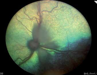
Feline fundus. Normal variation in optic disc myelination, with myelin radiating from the optic nerve head toward the tapetal region.
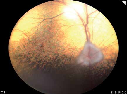
Canine fundus. Normal variation in optic disc myelination. Because of the greater amount of myelin, the optic nerve head is not sharply demarcated and has indistinct margins in some areas.
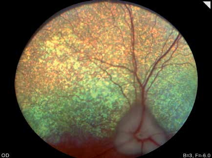
Canine fundus. Normal variation in optic disc myelination. A blurred margin is visible at the top of the optic nerve head.
Adapted from the Atlas of Ocular Diseases in Dogs and Cats by Dr. Siniša D. Grozdanić and colleagues. © Animal Eye Consultants of Iowa. The text and clinical images on this page are original content from the authors' own clinical practice and research, and may not be reproduced, distributed, or repurposed without permission.
Think your pet may have variations in optic nerve myelination?
Early evaluation gives us the most treatment options and the best chance of preserving vision.