
Uveodermatologic Syndrome (UVD)
Uveodermatologic syndrome is an immune-mediated disorder in which the body develops an immune response directed against melanocytes, resulting in depigmentation of skin and hair together with uveitis. Both eyes are usually affected, although severity may differ. Predisposed breeds listed in the source include Akitas, Siberian Huskies, Chow Chows, German Shepherd Dogs, Samoyeds, Shetland Sheepdogs, Rottweilers, and Golden Retrievers.
Ophthalmic manifestations include anterior uveitis, chorioretinitis, or panuveitis. Cataract, retinal detachment, iris bombe, hyphema, and glaucoma may develop. Characteristic dermatologic changes include depigmentation of skin and hair, with periocular and nasal vitiligo, which owners may notice first. Uveitis can also be the only initial manifestation and may precede skin changes.
Treatment
Diagnosis is based on the clinical presentation and histopathologic examination of representative tissue. Treatment requires long-term immunosuppression. Corticosteroids are administered topically and systemically, often with other immunosuppressive drugs such as cyclosporine. Clinical signs may improve rapidly after treatment begins but can recur if treatment is discontinued. Blindness caused by glaucoma or retinal detachment can occur despite therapy.
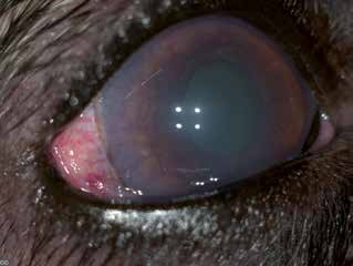
Chronic uveitis in a dog with uveodermatologic syndrome. Corneal vascularization and edema, altered iris appearance caused by posterior synechiae, and pronounced conjunctival and scleral vascularity are visible. High intraocular pressure was recorded in this dog.
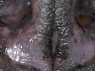
Uveodermatologic syndrome. The same dog as in the previous figure. Depigmentation of the nasal planum is visible.
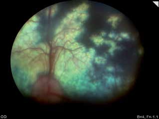
Uveodermatologic syndrome. Fundus appearance in the same dog. Retinal detachment and altered fundic pigmentation are visible.
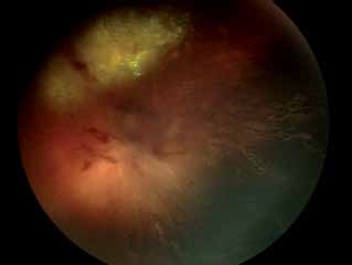
Uveodermatologic syndrome. Fundus of a dog with chronic chorioretinitis.
Adapted from the Atlas of Ocular Diseases in Dogs and Cats by Dr. Siniša D. Grozdanić and colleagues. © Animal Eye Consultants of Iowa. The text and clinical images on this page are original content from the authors' own clinical practice and research, and may not be reproduced, distributed, or repurposed without permission.
Think your pet may have uveodermatologic syndrome (uvd)?
Early evaluation gives us the most treatment options and the best chance of preserving vision.