
Uveal Melanoma and Melanocytoma
Uveal melanoma is a malignant tumor arising from uveal melanocytes and is among the most common primary intraocular neoplasms in dogs. It occurs mainly in older dogs. Uveal melanocytoma is benign but may be locally invasive. Anterior uveal melanocytomas of the iris or ciliary body, limbal melanocytomas, and choroidal melanocytomas are described.
Melanoma is most often located in the iris or ciliary body. Canine melanoma tends to grow nodularly rather than diffusely as in cats. It may appear as a tiny, barely visible pigmented lesion or as a large mass protruding through the pupil, displacing the iris anteriorly and producing dyscoria. Complications include uveitis, hyphema, secondary glaucoma, buphthalmos, retinal detachment, lens subluxation, and loss of vision.
Treatment
Diagnosis is based on ophthalmic examination, ultrasonography to help distinguish cyst from tumor, and gonioscopy to help distinguish uveal melanoma from limbal melanocytoma. A thorough systemic examination and CT or MRI are recommended because of metastatic potential. For uveal melanoma, the source recommends enucleation to reduce the risk of metastasis. Histopathologic examination is required to confirm the diagnosis.
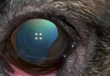
Uveal melanocytoma in a dog. The pupil has lost its normal shape.
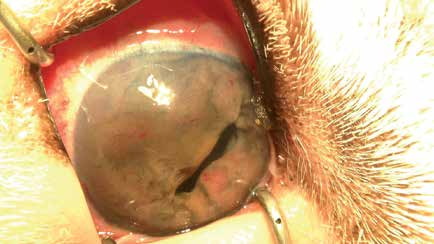
Iris melanoma, exceptionally advanced case. The pupil is barely visible.
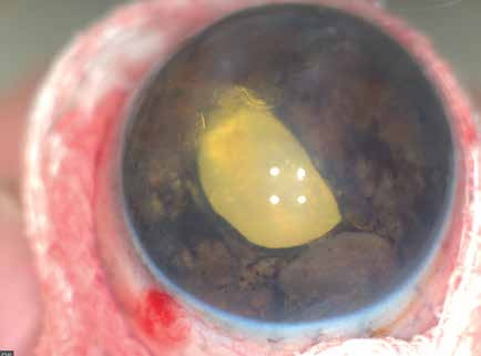
Iris melanoma that caused secondary glaucoma and vision loss in a dog. Globe after enucleation.
Adapted from the Atlas of Ocular Diseases in Dogs and Cats by Dr. Siniša D. Grozdanić and colleagues. © Animal Eye Consultants of Iowa. The text and clinical images on this page are original content from the authors' own clinical practice and research, and may not be reproduced, distributed, or repurposed without permission.
Think your pet may have uveal melanoma and melanocytoma?
Early evaluation gives us the most treatment options and the best chance of preserving vision.