
Trichiasis
Trichiasis occurs when normally positioned hairs of the periocular region come into contact with the cornea. Hairs reaching the ocular surface may originate from the eyelids, medial canthus, or nasal fold. Trichiasis may be unilateral or bilateral and may be congenital or acquired. Breeds predisposed to trichiasis include the Cocker Spaniel, Shih Tzu, Pug, and Miniature Poodle. It is common in breeds with excessive skin folds on the head, such as the Shar-Pei, Bulldog, Bloodhound, and Chow Chow. Acquired anatomical abnormalities, including eyelid lacerations or entropion, can also result in trichiasis.
The clinical presentation includes marked tearing (epiphora), eyelid spasm, and keratitis with or without ulceration. Just as entropion can cause trichiasis, pain from trichiasis and the resulting eyelid spasm can lead to secondary entropion.
Treatment
In some patients, regular trimming of periocular hair is sufficient. In others, permanent removal of individual hair follicles by cryoepilation is required. If entropion is the cause of trichiasis, surgical correction of entropion is recommended. In breeds with excessive skin folds on the head, excess skin can be surgically removed or temporary sutures can be placed to draw the eyelid skin away from the corneal surface, generally in puppies and young dogs. If hairs from the nasal fold contact the eye, surgical correction of that region is performed. If trichiasis results from an eyelid laceration, corrective eyelid surgery is necessary, with particular attention to proper apposition of the eyelid-margin edges. Consequences of trichiasis, including keratitis and corneal ulcers, must be treated.
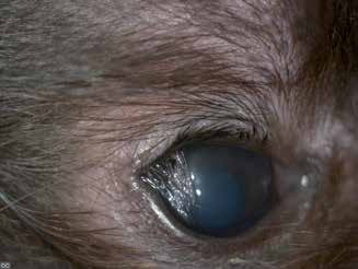
Trichiasis in a dog.
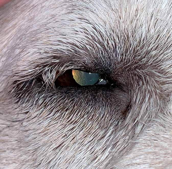
Trichiasis in a dog.
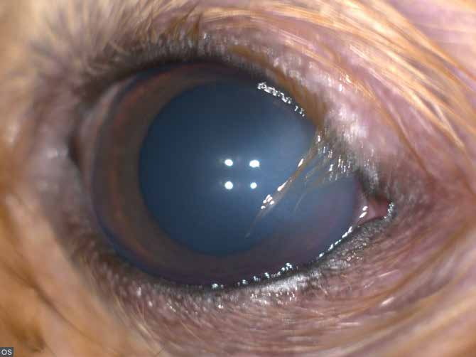
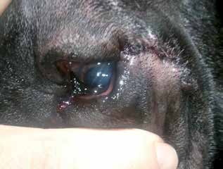
Trichiasis in a dog. Edema is present in the portion of the cornea contacting the hair (blue-gray corneal coloration).
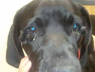
Dermoid of the lower eyelids in a young Great Dane with secondary corneal trichiasis.
Dermoid of the lower eyelids in a young Great Dane. The same dog as in the previous figure after surgical reconstruction and creation of a new eyelid margin, resolving the secondary trichiasis - left eye.
Adapted from the Atlas of Ocular Diseases in Dogs and Cats by Dr. Siniša D. Grozdanić and colleagues. © Animal Eye Consultants of Iowa. The text and clinical images on this page are original content from the authors' own clinical practice and research, and may not be reproduced, distributed, or repurposed without permission.
Think your pet may have trichiasis?
Early evaluation gives us the most treatment options and the best chance of preserving vision.