
Symblepharon
Symblepharon is adhesion of conjunctival tissue to another conjunctival surface and/or to the cornea. It most commonly develops as a consequence of herpesvirus and neonatal bacterial ocular and/or periocular infections in cats and occurs very rarely in dogs. Symblepharon usually develops secondary to severe corneal and conjunctival lesions. These lesions may result from chemical or mechanical injury to the conjunctiva and/or cornea, infectious disease such as herpesvirus or other viral and bacterial infections, or iatrogenic injury after surgery.
The eye may show corneal scarification and adhesion of conjunctiva to conjunctiva or to a superficial corneal layer that no longer has normal histologic structure. Conjunctival edema and pathological ocular discharge may be present. Adhesions narrow the palpebral opening and reduce eyelid mobility. If an adhesion is located near the opening of the nasolacrimal duct, impaired tear drainage results in epiphora.
Treatment
Symblepharon is identified during clinical examination. Therapy is directed toward the primary cause; inflammation is reduced with anti-inflammatory medications and viral and/or bacterial infection is treated. Existing adhesions are surgically removed. Surgically separated conjunctival surfaces, or conjunctiva and cornea, must be physically separated with a lens or silicone implants during healing. This is necessary to prevent formation of new adhesions, which is the most common complication of surgery.
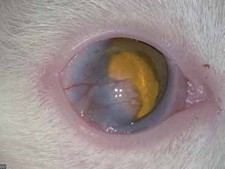
Symblepharon in a cat. Adhesion between the conjunctiva and cornea is pronounced in the lateral half of the eye. Conjunctival blood vessels are clearly visible.
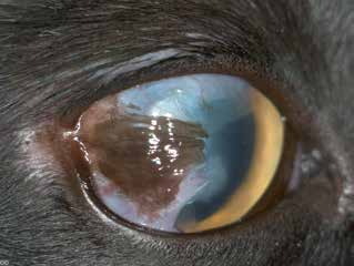
Symblepharon in a cat. A larger area of the eye is affected and pigment deposition is present. Only a small portion of the cornea near the medial canthus remains transparent.
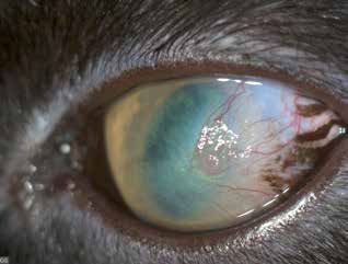
Symblepharon in a cat. In this case, the entire corneal surface is opaque. A network of blood vessels and tiger-stripe-like pigment deposits are visible.
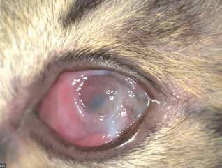
Symblepharon in a cat. Very severe case with adhesions between the conjunctiva of the third eyelid and the cornea. The cornea has completely lost transparency.
Adapted from the Atlas of Ocular Diseases in Dogs and Cats by Dr. Siniša D. Grozdanić and colleagues. © Animal Eye Consultants of Iowa. The text and clinical images on this page are original content from the authors' own clinical practice and research, and may not be reproduced, distributed, or repurposed without permission.
Think your pet may have symblepharon?
Early evaluation gives us the most treatment options and the best chance of preserving vision.