
Senile Iris Atrophy
Senile iris atrophy is progressive thinning of the iris stroma or pupillary margin and occurs in older dogs of all breeds. It may also occur in older cats. In Miniature Poodles, age-related atrophy of the iris sphincter can occur; as it progresses, the pupillary light reflex is initially absent and later the pupils become fixed and dilated, with anisocoria.
In many small dog breeds, stromal atrophy gradually leads to extensive loss of iris stroma and dyscoria. Regardless of the type of atrophy, focal iris color changes may be visible. These areas may initially appear hyperpigmented but lose pigment and become translucent as atrophy progresses. Senile iris atrophy is generally bilateral but not necessarily symmetric. Vision is not threatened, although photophobia may develop as the condition progresses.
Treatment
Diagnosis is established by ophthalmic examination. No treatment is performed.
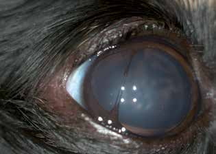
Pronounced iris atrophy in a dog. Most of the iris stroma has been lost, the pupil is dilated, and dyscoria is visible.
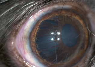
Iris atrophy in a dog. Iris tissue remains around the pupil, but much of the iris base is thinned or absent.
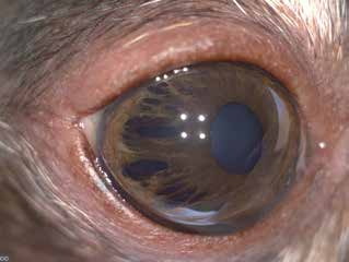
Iris atrophy in a dog. A similar but milder change than in the previous figure.
Adapted from the Atlas of Ocular Diseases in Dogs and Cats by Dr. Siniša D. Grozdanić and colleagues. © Animal Eye Consultants of Iowa. The text and clinical images on this page are original content from the authors' own clinical practice and research, and may not be reproduced, distributed, or repurposed without permission.
Think your pet may have senile iris atrophy?
Early evaluation gives us the most treatment options and the best chance of preserving vision.