
Secondary Glaucoma
Glaucoma consists of progressive ocular pathologic processes associated with increased intraocular pressure, visual impairment, and characteristic optic nerve changes. Reduced aqueous-humor outflow, increased intraocular pressure, reduced perfusion of the optic nerve head, structural optic nerve head changes, and neuroinflammation with retinal ganglion-cell abnormalities occur. High pressure may cause buphthalmos, stromal corneal edema, optic nerve anoxia, axonal death, and gliosis.
Secondary glaucoma develops because a preexisting ocular disease prevents normal aqueous-humor outflow. The underlying disorder may be a primary ocular disease or a systemic disease with ocular manifestations. Causes listed in the source include infectious diseases such as toxoplasmosis, neosporosis, bartonellosis, brucellosis, canine distemper, adenovirus infection, FIP, FeLV, blastomycosis, and cryptococcosis; metabolic disorders such as diabetes mellitus, systemic hypertension, and hyperlipidemia; neoplastic disease such as lymphoma, melanoma, myeloma, and sarcoma; immune-mediated disorders such as phacolytic and phacoclastic uveitis and uveodermatologic syndrome; idiopathic lymphoplasmacytic disorders; and blunt or penetrating ocular trauma.
Many of these conditions cause uveitis, and secondary glaucoma most commonly develops as a consequence of uveitis. Secondary glaucoma is also frequently associated with lens luxation, cataract, phacoemulsification, iris bombe, intraocular pigment deposition, retinal detachment, and numerous other ocular disorders. Clinical signs include elevated intraocular pressure, mydriasis or miosis, corneal edema with possible Haab's striae, secondary ulcerative keratitis, uveitis, episcleral vascular congestion, buphthalmos, lens subluxation, cortical cataract, vitreous degeneration, retinal degeneration, optic nerve head atrophy, and decreased vision or blindness. One or both eyes may be affected.
Treatment
Glaucoma is diagnosed by tonometry. Ocular ultrasonography can be used to evaluate intraocular structures when the cornea is opaque. A detailed ophthalmic examination may identify the primary disease responsible for glaucoma. If uveitis is present, its cause must be investigated. Glaucoma itself is not life-threatening, but infectious and neoplastic diseases can be, making identification and treatment of the primary disorder important. In addition to therapy directed at the primary disease, the source describes medications that reduce aqueous-humor production, such as carbonic anhydrase inhibitors and beta blockers, as safe choices in secondary glaucoma.
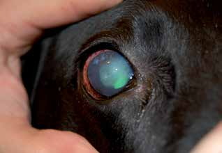
Secondary glaucoma following cataract surgery.
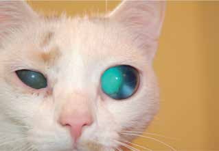
Secondary glaucoma in a cat due to uveitis. Buphthalmos, cataract, and lens luxation are visible.
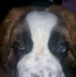
Secondary glaucoma after corneal perforation. Buphthalmos, corneal edema, and corneal scarring are visible.
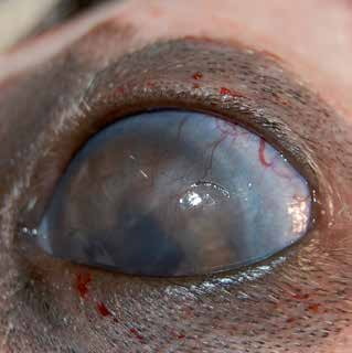
Secondary glaucoma in a dog caused by phacoclastic uveitis following a cat scratch and lens laceration.
Related in-depth guide
Glaucoma
Our full guide, including treatment and surgical options
Adapted from the Atlas of Ocular Diseases in Dogs and Cats by Dr. Siniša D. Grozdanić and colleagues. © Animal Eye Consultants of Iowa. The text and clinical images on this page are original content from the authors' own clinical practice and research, and may not be reproduced, distributed, or repurposed without permission.
Think your pet may have secondary glaucoma?
Early evaluation gives us the most treatment options and the best chance of preserving vision.