
Retrobulbar Neoplasia
Retrobulbar neoplasms are most commonly malignant tumors. They may develop within retrobulbar tissues or may represent extension of tumors from surrounding ocular tissues, the nasal cavity, or the oral cavity into the retrobulbar region. The most common neoplasms listed in the source include carcinomas such as squamous cell carcinoma and adenocarcinoma; sarcomas such as fibrosarcoma, hemangiosarcoma, multilobular orbital sarcoma, osteosarcoma, and chondrosarcoma; as well as meningioma and lymphoma.
Clinical findings may include periocular swelling, exophthalmos, conjunctival hyperemia and chemosis, protrusion of the third eyelid, ocular discharge, lagophthalmos, and keratitis. These tumors generally occur in older dogs and cats. The location of the mass can sometimes be inferred from the position of the globe; for example, a medially located mass may displace the globe laterally. Acute pain may develop if an abscess forms within the tumor, in which case the patient may react painfully during retropulsion or opening of the jaw.
Treatment
Retrobulbar neoplasia is detected using ultrasonography, radiography, CT, and MRI. Biopsy and fine-needle aspiration may assist in diagnosis, while the most reliable diagnosis is established by tumor excision and histopathologic examination. If possible, only the neoplasm is surgically removed, with radiotherapy and/or chemotherapy as needed; alternatively, enucleation or exenteration may be performed.
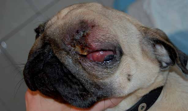
Retrobulbar neoplasia in a dog. Periocular swelling and exophthalmos are present.
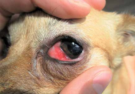
Retrobulbar neoplasia in a dog. Periocular swelling and altered pupil shape and position are present. The pupil is displaced laterally by the tumor because the entire globe is pushed forward and laterally.
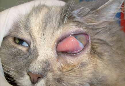
Retrobulbar neoplasia in a cat. Exophthalmos, protrusion of the third eyelid, lagophthalmos, and a corneal ulcer are present.
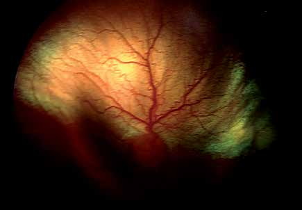
Funduscopic changes. Because the retrobulbar neoplasm compresses the globe from behind, deformation of the fundus can be seen.
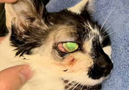
Retrobulbar neoplasia. This 12-year-old cat had marked exophthalmos, lagophthalmos, limited retropulsion, and corneal erosion.
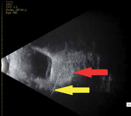
Retrobulbar neoplasia. Same cat as in the previous figure, ultrasonographic findings. A mass compressing and deforming the globe is visible (red arrow). The yellow arrow shows the needle used for ultrasound-guided fine-needle aspiration (UGFNA) for cytology. Cytology was suspicious for squamous cell carcinoma (SCC).
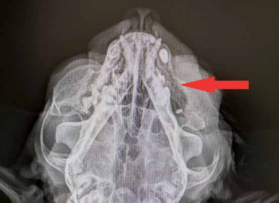
Radiograph of the head of the cat from the previous figure. Osteolysis of the maxilla is present (red arrow).
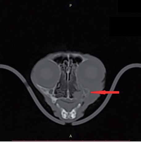
CT image of the head of the cat from the previous figures. The invasiveness of the lesion and osteolysis are clearly visible (red arrow). Note how the retrobulbar mass displaces the globe and produces exophthalmos.
Adapted from the Atlas of Ocular Diseases in Dogs and Cats by Dr. Siniša D. Grozdanić and colleagues. © Animal Eye Consultants of Iowa. The text and clinical images on this page are original content from the authors' own clinical practice and research, and may not be reproduced, distributed, or repurposed without permission.
Think your pet may have retrobulbar neoplasia?
Early evaluation gives us the most treatment options and the best chance of preserving vision.