
Retinal Hemorrhage
Retinal hemorrhage may occur within the retina or in the space in front of or behind it. It may result from primary retinal disease or systemic disease that predisposes to bleeding. Hemorrhages may be focal or generalized, limited to the retina or present in other ocular structures, and unilateral or bilateral.
Causes include congenital ocular disease, physical trauma, hypertension, diabetes mellitus, neoplasia, infectious disease, intoxication, coagulopathies, and vasculopathies. The source lists hypertension; tick-borne, fungal, bacterial, and viral infections; thrombocytopenia; anemia and pancytopenia caused by infectious or autoimmune disease; hyperviscosity syndrome; diabetes; and anticoagulant rodenticide intoxication among common causes in dogs and cats. Visual impairment ranges from mild to complete blindness depending on cause, extent of hemorrhage, and resulting ocular injury. Other clinical signs may include hyphema, petechial mucosal hemorrhage, or blood in the stool, depending on the primary disease.
Treatment
Retinal hemorrhage is readily identified by indirect ophthalmoscopy, but determining the underlying cause can be demanding. Diagnostic investigation may include ocular ultrasonography, complete blood count, serum biochemistry, urinalysis, blood-pressure measurement, thyroid and adrenal testing, coagulation testing, thoracic radiography, echocardiography, abdominal ultrasonography, and infectious-disease testing. Successful treatment of the primary disorder is important to stop further bleeding and prevent retinal detachment. Complete retinal detachment may require vitreoretinal surgery even after the primary disorder is controlled.
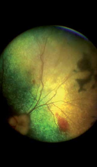
Retinal hemorrhages in a patient with autoimmune thrombocytopenia. Dark regions are old choroidal scars from previous retinal inflammation. Coagulopathies of different causes can produce retinal hemorrhage and, when severe, lead to retinal detachment.
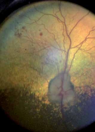
Focal retinal hemorrhages in a dog with confirmed rickettsial infection and clinical thrombocytopenia.
Related in-depth guide
Retinal Disorders
Our full guide, including treatment and surgical options
Adapted from the Atlas of Ocular Diseases in Dogs and Cats by Dr. Siniša D. Grozdanić and colleagues. © Animal Eye Consultants of Iowa. The text and clinical images on this page are original content from the authors' own clinical practice and research, and may not be reproduced, distributed, or repurposed without permission.
Think your pet may have retinal hemorrhage?
Early evaluation gives us the most treatment options and the best chance of preserving vision.