
Retinal Dysplasia (RD)
Retinal dysplasia is abnormal retinal differentiation resulting in multifocal or diffuse disorganization of retinal layers. This produces separation or abnormal apposition of the neuroretina and retinal pigment epithelium. In dogs, retinal dysplasia is most commonly hereditary and, depending on breed, may be inherited in an autosomal recessive or dominant manner. It can occur alone or with other ocular abnormalities. Less commonly, adverse influences during intrauterine retinal development, such as viral disease, radiation exposure, nutritional deficiency, or trauma, may contribute.
Fundic examination may reveal multiple V- or Y-shaped retinal folds or irregular rosettes that are dark gray or whitish, representing focal or multifocal retinal dysplasia. Changes are most commonly located in the central tapetal fundus. More severe disease may produce confluent, dark-bordered horseshoe-shaped lesions, termed geographic dysplasia. The most severe form is associated with retinal detachment, generalized dysplasia.
Vision is preserved in mild retinal dysplasia. Geographic dysplasia may cause visual deficits, while complete retinal dysplasia with retinal detachment results in blindness. Young dogs may occasionally show transient retinal folds because the retina develops faster than surrounding choroid and sclera; these can resemble dysplasia but are temporary and are not considered pathologic.
Treatment
Mild dysplasia that does not affect vision is often discovered incidentally during an examination for another problem or during certification. If suspected, fundic examination can identify the condition at six to eight weeks of age, but examination should be repeated between six and twelve months. Genetic tests exist for some breeds. No treatment is performed, and affected animals should be excluded from breeding according to the source.
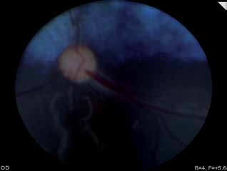
Retinal dysplasia in a young dog. Retinal folds appear as whitish lines in the nontapetal region.
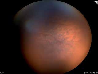
Multifocal retinal dysplasia in a young dog with focal lesions in the tapetal fundus.
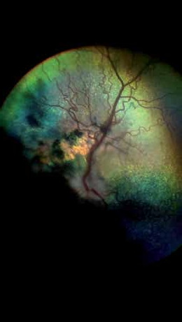
Geographic retinal dysplasia in a dog. Areas of hypo- and hyperreflectivity and tortuous retinal vessels are visible on fundic examination.
Related in-depth guide
Retinal Disorders
Our full guide, including treatment and surgical options
Adapted from the Atlas of Ocular Diseases in Dogs and Cats by Dr. Siniša D. Grozdanić and colleagues. © Animal Eye Consultants of Iowa. The text and clinical images on this page are original content from the authors' own clinical practice and research, and may not be reproduced, distributed, or repurposed without permission.
Think your pet may have retinal dysplasia (rd)?
Early evaluation gives us the most treatment options and the best chance of preserving vision.