
Progressive Retinal Atrophy (PRA)
Progressive retinal atrophy is a term for a group of inherited dystrophic or degenerative retinal diseases that cause dysfunction and death of photoreceptors. Different genetic mutations have been identified in different breeds, most often with autosomal recessive inheritance. Disease is always bilateral. Most forms begin with poor night vision because rods are affected first, although less commonly disease begins with impaired daylight vision because of primary cone degeneration. As disease progresses, vision is lost under both night and daylight conditions.
Early fundic changes may include altered reflectivity of the tapetal region and mild attenuation of retinal vessels. Later, hyperreflective areas gradually involve the entire tapetum, leaving only faint retinal vessel contours centrally. The nontapetal region develops irregular pigmentation with depigmented areas and pigment clumping. The optic disc becomes pale and loses myelination. The pupillary light reflex weakens and the pupils eventually remain dilated.
Age of onset depends on the mutation. Some forms present soon after birth, with poor night vision in the first weeks and complete blindness by approximately one year, as described for rod-cone dysplasia in Irish Setters, Collies, Corgis, Pit Bull Terriers, and Dachshunds. Other forms present later, with night-vision impairment between three and five years and complete blindness between five and seven years, such as classic PRA described in Poodles, Maltese, and Cocker Spaniels. In some breeds, disease begins in the retinal pigment epithelium and secondarily affects the neuroretina, as in RPED described in Labrador and Golden Retrievers, Border Collies, and Briards.
Treatment
Diagnosis is based on ophthalmic examination. Depending on the mutation, fundic changes may be visible as early as three to four months or not until years later; ERG abnormalities follow similarly variable timing. Genetic tests exist for some breeds. No specific treatment is described, and appropriate selection of breeding animals is emphasized.
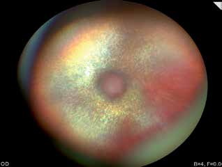
Progressive retinal atrophy in a cat. This advanced case shows loss of visible retinal vessels and prominent hyperreflective fundic areas.
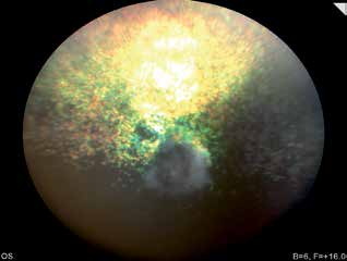
Progressive retinal atrophy in a dog. The optic nerve head is pale gray, retinal vessels are not visible, and hyperreflective areas are present.
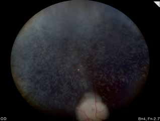
Progressive retinal atrophy in a dog. Thin, reduced retinal vessels demonstrate vascular attenuation.
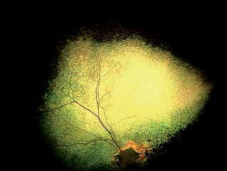
Progressive retinal atrophy in a dog. Hyperreflective areas and thin, attenuated retinal vessels are readily visible.
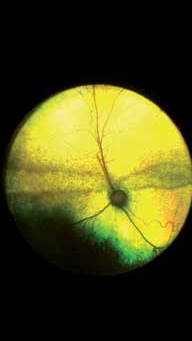
Central retinal degeneration in a cat. The source describes this type of degeneration as most commonly associated with inherited feline disease such as progressive retinal atrophy.
Related in-depth guide
Retinal Disorders
Our full guide, including treatment and surgical options
Adapted from the Atlas of Ocular Diseases in Dogs and Cats by Dr. Siniša D. Grozdanić and colleagues. © Animal Eye Consultants of Iowa. The text and clinical images on this page are original content from the authors' own clinical practice and research, and may not be reproduced, distributed, or repurposed without permission.
Think your pet may have progressive retinal atrophy (pra)?
Early evaluation gives us the most treatment options and the best chance of preserving vision.