
Primary Glaucoma
Glaucoma is a group of progressive ocular pathologic processes associated with elevated intraocular pressure, visual deterioration, and characteristic changes in the optic nerve. It involves reduced aqueous-humor outflow, increased intraocular pressure, reduced blood supply to the optic nerve head, structural optic nerve head changes, and neuroinflammation with abnormalities of retinal ganglion cells. High intraocular pressure can produce buphthalmos because of scleral elasticity, especially in young animals and in adults with chronic glaucoma. The cornea becomes thickened and cloudy because of stromal edema. Optic nerve anoxia, axonal death, and gliosis of the optic nerve head follow.
Primary glaucoma is hereditary glaucoma associated with mutations in different genes, with particular mutations characteristic of specific breeds. Breeds described as predisposed include Basset Hounds, Beagles, Cocker Spaniels, Siberian Huskies, Boston Terriers, Chow Chows, and Miniature Poodles. Clinical signs may occur in young dogs or may not become evident until approximately four, six, or even twelve years of age.
Dogs with primary glaucoma may have an open or closed iridocorneal angle, assessed by gonioscopy. The outflow pathway beyond the angle, including the ciliary cleft, should also be evaluated, for example with high-frequency ultrasonography, when determining whether primary glaucoma is present. Both eyes are affected, although disease may not be symmetric. When glaucoma is diagnosed in one eye, additional testing should determine whether it is primary or secondary because that distinction influences treatment of both the affected eye and the apparently healthy fellow eye.
Clinical signs include increased intraocular pressure, mydriasis, corneal edema with possible Haab's striae, secondary ulcerative keratitis, episcleral vascular congestion, buphthalmos, lens subluxation, secondary uveitis, cortical cataract, vitreous degeneration, retinal degeneration, and optic nerve head atrophy.
Treatment
Diagnosis is based on tonometry, gonioscopy, and high-frequency ultrasonography. When glaucoma is identified in one eye, the fellow eye should also be examined and prophylactic therapy of the fellow eye is initiated according to the source because it is considered at risk of future elevation of intraocular pressure. Medical therapy consists of antiglaucoma eye drops. Surgical procedures include transscleral or endoscopic laser cyclophotocoagulation to reduce aqueous-humor production and placement of various anterior chamber shunts to facilitate aqueous-humor drainage; these procedures are often combined.
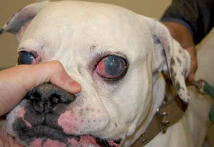
Primary glaucoma in an American Bulldog. Corneal edema, scleral and conjunctival hyperemia, and buphthalmos are visible. The dog is blind in both eyes.
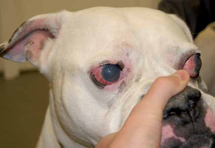
Primary glaucoma in a dog. The same dog as in the previous figure, opposite eye.
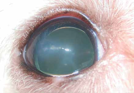
Primary glaucoma in a Mexican Hairless Dog. Buphthalmos, corneal edema, and lens luxation/subluxation are visible.
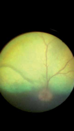
Retinal edema and a temporal area of retinal and choroidal infarction in a dog with acute primary hereditary glaucoma.
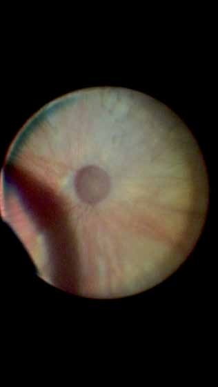
Optic nerve and retinal damage caused by high intraocular pressure in a dog with primary glaucoma.
Related in-depth guide
Glaucoma
Our full guide, including treatment and surgical options
Adapted from the Atlas of Ocular Diseases in Dogs and Cats by Dr. Siniša D. Grozdanić and colleagues. © Animal Eye Consultants of Iowa. The text and clinical images on this page are original content from the authors' own clinical practice and research, and may not be reproduced, distributed, or repurposed without permission.
Think your pet may have primary glaucoma?
Early evaluation gives us the most treatment options and the best chance of preserving vision.