
Posterior Lens Luxation
The lens may dislocate anteriorly into the anterior chamber or posteriorly into the vitreous. As described for anterior luxation, primary bilateral hereditary lens luxation is associated in the source with an ADAMTS gene mutation and occurs particularly in terriers, Jack Russell Terriers, Border Collies, Chow Chows, and related mixed breeds, usually between three and six years of age. The process is bilateral but not symmetric. Secondary luxation may occur with abnormal ocular development, degeneration, trauma, uveitis, cataract, or glaucoma.
Posterior lens luxation causes less acute pain than anterior luxation. Vitreous degeneration is a prerequisite for posterior dislocation. The source states that a lens lying on the retinal surface causes tractional retinal detachment and blindness, a process that may develop over months to years. A dislocated lens may change position within the vitreous or may move from a posterior to an anterior location.
Treatment
Diagnosis is made by ophthalmic examination, with visualization of the lens margin and a crescent-shaped aphakic reflection caused by increased tapetal reflection. Vitreous degeneration appears as floating deposits within the normally transparent vitreous. Ultrasonography can confirm the diagnosis. Miotics may be used long-term to retain the lens in the posterior segment together with anti-inflammatory medications. Because the source states that posterior lens luxation causes retinal detachment, surgical lens removal combined with vitrectomy and silicone-oil retinal tamponade is generally indicated. If the lens moves anteriorly, intracapsular extraction or phacoemulsification is recommended.
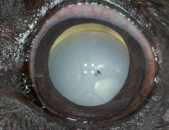
Posteriorly subluxated cataractous lens in a dog. The aphakic crescent is clearly visible.
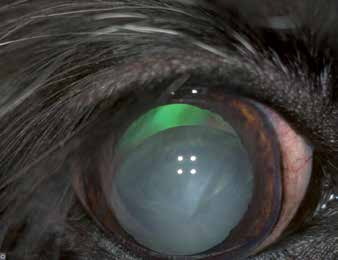
Posteriorly subluxated cataractous lens in a dog.
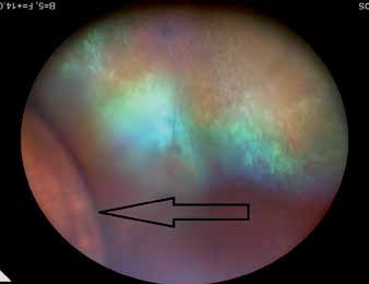
Posterior lens luxation. The black arrow indicates the lens equator lying on the retinal surface. The fundus is visible behind the lens.
Adapted from the Atlas of Ocular Diseases in Dogs and Cats by Dr. Siniša D. Grozdanić and colleagues. © Animal Eye Consultants of Iowa. The text and clinical images on this page are original content from the authors' own clinical practice and research, and may not be reproduced, distributed, or repurposed without permission.
Think your pet may have posterior lens luxation?
Early evaluation gives us the most treatment options and the best chance of preserving vision.