
Persistent Pupillary Membranes (PPM)
Persistent pupillary membranes are remnants of embryonic vasculature and associated connective tissue that supplied the lens with nutrients during intrauterine development. Complete regression should normally be completed by approximately six weeks of age, although in some dogs the process can continue for several months after birth.
If resorption is incomplete, persistent pupillary membranes remain. They appear as fine pigmented strands, light to dark brown, that always originate from the iris. Membranes may extend from one region of iris to another on the same side of the pupil, across the pupil, from iris to corneal endothelium where they can cause corneal opacity, from iris to lens where they may produce cataract, or they may float freely in the anterior chamber.
PPMs generally do not cause significant visual problems except in uncommon cases in which they produce corneal opacity or secondary cataract, which is usually nonprogressive. In some dog breeds, including Basenjis, Welsh Corgis, Mastiffs, Chow Chows, and English Cocker Spaniels, PPM is considered hereditary. Because PPM can accompany other ocular abnormalities, the source recommends against breeding animals with this finding. In cats, upper eyelid agenesis is often associated with PPM.
Treatment
Diagnosis is made by ophthalmic examination. Treatment is rarely necessary. If adhesions to the corneal endothelium and secondary corneal edema are present, topical hypertonic sodium chloride, such as 5% NaCl ophthalmic ointment, may be used. Cataract surgery is performed if necessary. Surgical excision of the membranes is rarely required.
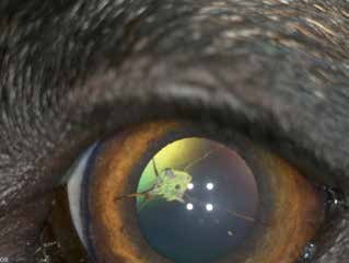
Persistent pupillary membranes in a dog extending from the iris to the cornea. A rhomboid area of corneal opacity is visible where the membranes contact the corneal endothelium.
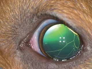
Persistent pupillary membrane in a dog extending from one side of the iris to the other.
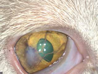
Persistent pupillary membranes extending from iris to cornea. Corneal fibrosis and edema are visible at the site of contact in this cat.
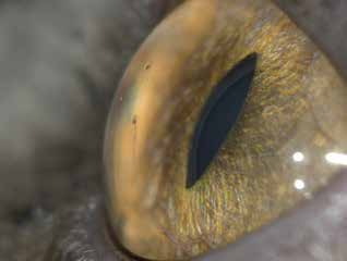
Persistent pupillary membranes in a cat, appearing as fine cobweb-like strands extending from iris to cornea. Because the eye is photographed from the side, the point at which the membranes meet the corneal endothelium is clearly visible.
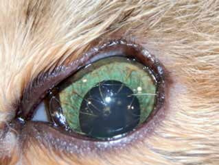
Persistent pupillary membranes in a cat extending from iris to lens, with agenesis of the lateral half of the upper eyelid as an associated genetic anomaly.
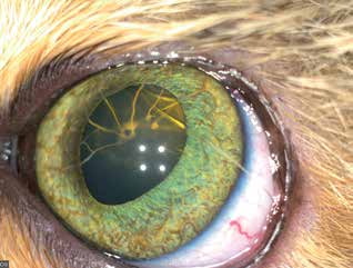
Persistent pupillary membranes in the same cat as in the previous figure, photographed from another angle. Upper eyelid agenesis is less obvious in this view, but localized opacities on the anterior lens capsule where the membranes contact the lens are easier to see.
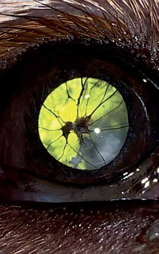
Persistent pupillary membranes in a dog extending from iris to lens. Lens opacity is visible where the membranes contact the lens surface because of cataract formation.
Adapted from the Atlas of Ocular Diseases in Dogs and Cats by Dr. Siniša D. Grozdanić and colleagues. © Animal Eye Consultants of Iowa. The text and clinical images on this page are original content from the authors' own clinical practice and research, and may not be reproduced, distributed, or repurposed without permission.
Think your pet may have persistent pupillary membranes (ppm)?
Early evaluation gives us the most treatment options and the best chance of preserving vision.