
Persistent Hyaloid Vasculature (PHV)
During embryonic development, the lens receives its blood supply from the intravitreal hyaloid vascular system. Regression of this system begins around day 45 of gestation. In some dog breeds, remnants of hyaloid vasculature persist, often together with other ocular abnormalities such as persistent pupillary membranes, microphthalmia, microphakia, lens coloboma, elongated ciliary processes, and cataract.
The source describes the anomaly as autosomal recessive and bilateral, most commonly in Doberman Pinschers, Schnauzers, and Staffordshire Bull Terriers, and less commonly in other breeds. It may also occur sporadically as a nonhereditary unilateral anomaly. Clinically, retrolenticular or intralenticular pigmented spots or deposits, blood or blood clots, and persistent portions of the hyaloid system may be visible. The posterior lens capsule may be affected and can rupture. Secondary cataract is common. Other possible complications include hyphema, uveitis, and secondary glaucoma.
Treatment
Diagnosis is made by ophthalmic examination and may be aided by color Doppler ultrasonography. When necessary, treatment is surgical and consists of phacoemulsification with posterior capsulectomy and intraocular lens implantation. Severely affected animals are excluded from breeding according to the source.
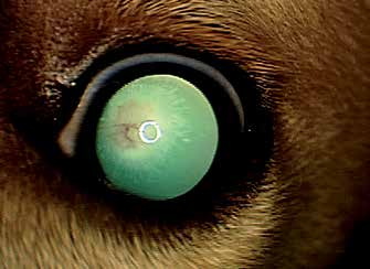
Persistent hyaloid vasculature in a dog. Localized opacity of the posterior lens capsule and blood-containing persistent vessels of the tunica vasculosa lentis are visible.
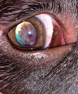
Persistent hyaloid vasculature in a dog. Localized opacity of the posterior lens capsule caused by cataract and persistent blood-containing vessels of the tunica vasculosa lentis are visible.
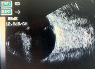
Ultrasound image of the same dog. A vessel extending from the optic nerve surface toward the posterior lens capsule is visible.
Adapted from the Atlas of Ocular Diseases in Dogs and Cats by Dr. Siniša D. Grozdanić and colleagues. © Animal Eye Consultants of Iowa. The text and clinical images on this page are original content from the authors' own clinical practice and research, and may not be reproduced, distributed, or repurposed without permission.
Think your pet may have persistent hyaloid vasculature (phv)?
Early evaluation gives us the most treatment options and the best chance of preserving vision.