
Palpebral Melanoma (Eyelid Melanoma)
Eyelid melanoma is a malignant neoplasm arising from melanocytes. This tumor behaves invasively, with a high recurrence rate and marked metastatic potential. It may appear as a firm, irregularly shaped, darkly pigmented mass or, occasionally, as a pale lesion. Melanoma is most commonly located directly on the eyelid margin at the mucocutaneous junction.
Two types of melanoma are recognized: melanotic, which contains melanin, and amelanotic, which lacks melanin. Histopathologic diagnosis of the amelanotic form is often difficult, and final diagnosis is obtained by immunocytochemical analysis. Older dogs are most commonly affected, with a mean age of nine years in the source. Predisposed breeds include the Airedale Terrier, Boston Terrier, Boxer, Chihuahua, Chow Chow, Cocker Spaniel, Poodle, Rottweiler, and Labrador Retriever. Eyelid melanoma is rarely diagnosed in cats.
Treatment
Diagnosis is established by histopathologic examination and, for amelanotic melanoma, immunocytochemical analysis. Therapy is carefully selected according to the stage of disease. Stages I-IV are determined based on tumor size and the presence of metastases. The source recommends surgical removal of the tumor, together with radiation therapy, chemotherapy, and immunotherapy; melanoma vaccines have been in use since 2007.
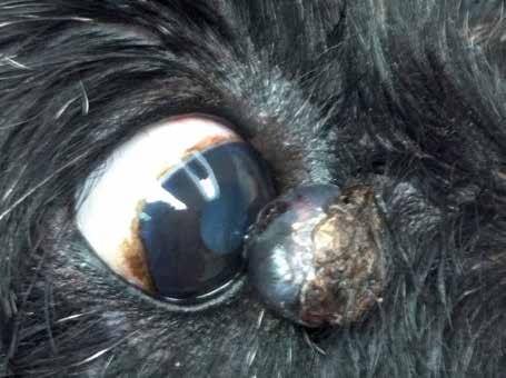
Large melanoma at the medial canthus in a dog, contacting the cornea.
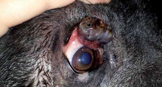
Upper eyelid melanoma in a Shar-Pei causing marked ocular irritation.
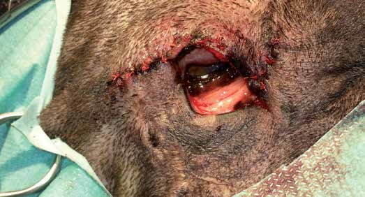
Upper eyelid melanoma in a Shar-Pei. Tumor resection with eyelid reconstruction using a split-lid technique.
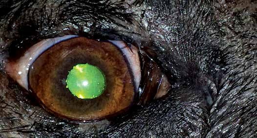
Upper eyelid melanoma in a Shar-Pei - postoperative photograph two months after surgery.
Adapted from the Atlas of Ocular Diseases in Dogs and Cats by Dr. Siniša D. Grozdanić and colleagues. © Animal Eye Consultants of Iowa. The text and clinical images on this page are original content from the authors' own clinical practice and research, and may not be reproduced, distributed, or repurposed without permission.
Think your pet may have palpebral melanoma (eyelid melanoma)?
Early evaluation gives us the most treatment options and the best chance of preserving vision.