
Palpebral Melanocytoma (Eyelid Melanocytoma)
Eyelid melanocytoma is a benign tumor of melanocytes. It appears as a solitary or multiple cutaneous lesion that is small, slow-growing, irregular in shape, and most commonly hyperpigmented, brown, or almost black. The cells composing the tumor may vary considerably in morphology. Several cell types may be present, but the cytoplasm generally contains variable quantities of pigment. Some cells may be completely devoid of melanin, which makes diagnosis more difficult. The tumor occurs in dogs and is extremely rare in cats. It can occur in all dog breeds but has been reported somewhat more commonly in Vizslas, Miniature Schnauzers, Doberman Pinschers, and Airedale Terriers.
Treatment
An accurate diagnosis is very important because melanocytoma and melanoma may appear morphologically similar, although one is benign and the other malignant. Diagnosis is established by histopathologic examination. If the lesions cause no problems, they may be left in place and monitored. Otherwise, they can be removed by conventional surgery, cryosurgery, or laser. Recurrence is possible, as is the development of new melanocytomas at other locations.
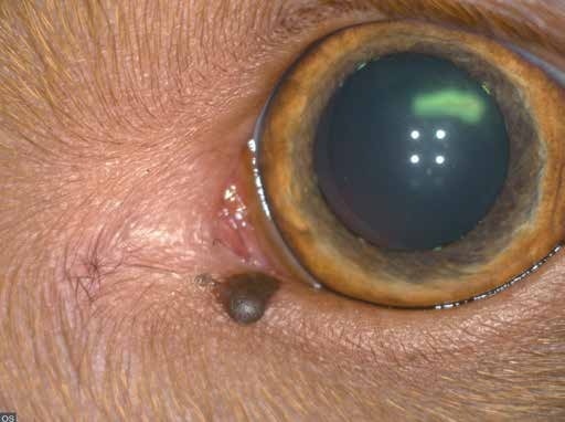
Melanocytoma of the lower eyelid near the medial canthus.
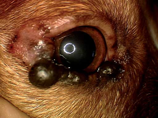
Multiple melanocytomas of the lower eyelid in a dog.
Adapted from the Atlas of Ocular Diseases in Dogs and Cats by Dr. Siniša D. Grozdanić and colleagues. © Animal Eye Consultants of Iowa. The text and clinical images on this page are original content from the authors' own clinical practice and research, and may not be reproduced, distributed, or repurposed without permission.
Think your pet may have palpebral melanocytoma (eyelid melanocytoma)?
Early evaluation gives us the most treatment options and the best chance of preserving vision.