
Orbital Cellulitis and Orbital Abscess
Orbital cellulitis is inflammation of the soft tissues of the orbital space. A common cause is a sharp foreign body penetrating through the oral cavity behind the last molar, through the conjunctiva, or through the eyelids and entering the orbit. Cellulitis may also result from systemic infections or extension of infection from nearby structures, including salivary glands, the nasal cavity or sinuses, and the oral cavity, particularly the teeth. Endophthalmitis and panophthalmitis may also lead to orbital cellulitis.
An abscess may frequently form. Bacteria isolated most commonly from abscesses in the source include Pasteurella spp., E. coli, Staphylococcus spp., and Bacteroides. Orbital abscesses are often associated with vasculitis, or inflammation of blood-vessel walls. As a consequence of vasculitis and increased vascular permeability, bacteria can migrate from any part of the body, most commonly from inflamed skin, into the orbital space and lead to formation of an orbital abscess. Vasculitis can be confirmed by histopathologic analysis of orbital soft-tissue samples. Persistent medical control of vasculitis is a very important component of abscess treatment because abscess recurrence has been observed when vasculitis is not controlled. Fungal or parasitic infections may occasionally cause cellulitis.
The clinical presentation most commonly includes acute unilateral exophthalmos with protrusion of the third eyelid, conjunctival hyperemia and chemosis, episcleral congestion, and mucopurulent ocular discharge. Periocular swelling varies in severity, and marked pain is present when the jaw is opened.
Treatment
Diagnosis is made through detailed clinical examination supported by radiography, which may identify a metallic foreign body or bone fracture, ultrasonography, and computed tomography. Any foreign body must be removed and an abscess drained, either through the oral cavity or by a retrobulbar approach. Local antibiotic irrigation, systemic antibiotics, and nonsteroidal anti-inflammatory medications are used.
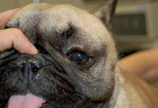
Orbital cellulitis in a dog that developed after a bite sustained during a fight with another dog.

Orbital cellulitis in a dog after penetration of a foreign body from the oral cavity into the retrobulbar space.
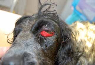
Orbital cellulitis in a dog caused by a foreign body.
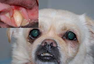
Orbital cellulitis in a dog caused by infection of a maxillary tooth. The problematic tooth and purulent material around it can be seen in the upper left corner.
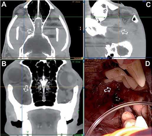
Orbital abscess in a dog associated with orbital vasculitis. A) CT showed an orbital abscess at the base of the orbit (white arrow), inflammatory changes in the orbital soft tissues, and exophthalmos. B) CT showed swelling of orbital soft tissues, but the abscess itself was not clearly visible. C) CT showed the orbital abscess (white arrow). A linear shadow within the abscess is often seen with a foreign body, but in this case it was an imaging artifact. The abscess lies below the axial zone of the globe, which is very likely why orbital ultrasonography failed to identify it in this case. D) Soft-tissue swelling behind the last molar is a typical finding with an orbital abscess. Opening the mouth is very painful in these dogs; therefore, this examination should be performed under general anesthesia with endotracheal intubation. Surgical resection of the orbital floor may result in substantial drainage of blood and purulent material, and aspiration of this material can cause pneumonia.
Adapted from the Atlas of Ocular Diseases in Dogs and Cats by Dr. Siniša D. Grozdanić and colleagues. © Animal Eye Consultants of Iowa. The text and clinical images on this page are original content from the authors' own clinical practice and research, and may not be reproduced, distributed, or repurposed without permission.
Think your pet may have orbital cellulitis and orbital abscess?
Early evaluation gives us the most treatment options and the best chance of preserving vision.