
Optic Neuritis
Inflammation of the optic nerve may be unilateral but is most commonly bilateral. The optic nerve head and/or the retrobulbar portion of the nerve may be affected. Causes listed in the source include autoimmune disease, either isolated immune-mediated optic neuritis or optic neuritis associated with meningoencephalitis; infectious diseases such as canine distemper, leptospirosis, borreliosis, blastomycosis, cryptococcosis, histoplasmosis, neosporosis, ehrlichiosis, and toxoplasmosis; and neoplastic disease, either direct involvement of the optic nerve or an immune reaction associated with cancer.
The source emphasizes that optic neuritis may be the only initial sign of meningoencephalitis of unknown etiology (MUE), with other neurologic signs developing considerably later. Less common causes include trauma, toxins, or extension of inflammatory or neoplastic disease from surrounding ocular structures. In many cases, no cause is identified and the condition is considered idiopathic.
The optic disc may be edematous with indistinct blurred margins. Blood vessels crossing the disc may be elevated and hemorrhage may be present. Peripapillary edema and neuroretinitis may also occur. Blindness develops suddenly and is accompanied by dilated pupils that do not respond to light. If meningitis is also present, other neurologic signs may be observed. The ERG is generally without significant abnormality.
Treatment
Optic neuritis can resemble papilledema associated with optic nerve compression from extraocular myositis or intracranial hypertension, so these disorders must be differentiated. A detailed ophthalmic and general clinical examination, complete blood count and serum biochemistry, MRI, and infectious-disease testing can assist in determining the cause. Cerebrospinal fluid analysis may be useful when meningoencephalitis is suspected. Treatment is directed at the primary cause and includes corticosteroids and other immunosuppressive medications. Prognosis for recovery of vision depends on the primary disease and the duration of blindness before treatment begins.
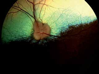
Optic neuritis in a dog without obvious optic nerve swelling. Optic neuritis was suspected on the basis of the patient's clinical signs.
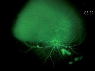
Optic neuritis in the same dog as in the previous figure after fluorescein angiography. Chorioretinal exudative lesions become visible with this procedure, including an exudative lesion at the optic nerve head.
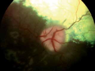
Optic neuritis with sudden loss of vision secondary to granulomatous meningoencephalitis. Papilledema and altered direction of blood vessels at the margin of the optic disc are visible. Whitish lesions in the ventral fundus are exudative lesions often seen in dogs with optic neuritis.

Optic neuritis. The same dog whose fundus is shown in the previous figure. Bilateral mydriasis is visible.
Adapted from the Atlas of Ocular Diseases in Dogs and Cats by Dr. Siniša D. Grozdanić and colleagues. © Animal Eye Consultants of Iowa. The text and clinical images on this page are original content from the authors' own clinical practice and research, and may not be reproduced, distributed, or repurposed without permission.
Think your pet may have optic neuritis?
Early evaluation gives us the most treatment options and the best chance of preserving vision.