
Optic Nerve Hypoplasia/Aplasia/Coloboma
Optic nerve hypoplasia is characterized by a small optic nerve head diameter caused by a reduced number of axons. Two terms are commonly used to grade this anomaly. Micropapilla refers to an optic nerve head smaller than normal with fewer axons but without impaired vision or pupillary light reflexes. Optic nerve hypoplasia is used when the axon count is sufficiently reduced to cause visual deficits and abnormal pupillary reflexes. These anomalies may result from abnormal ganglion-cell development or premature ganglion-cell death.
Clinically, the optic nerve head is small, gray, and lacks myelination. In hypoplasia, the pupils are dilated and nonresponsive. Hereditary optic nerve hypoplasia has been reported in Miniature Poodles, Collies, Shetland Sheepdogs, Saint Bernards, and Beagles, and less commonly in other breeds.
Optic nerve aplasia is complete absence of retinal ganglion cells and the optic nerve. Retinal vessels are absent. The source describes aplasia in Shih Tzus; affected dogs are blind.
Optic nerve coloboma is a congenital, developmental, nonprogressive malformation involving the optic nerve head and peripapillary retina. It may or may not impair vision; severe coloboma can cause blindness. Indirect ophthalmoscopy may reveal an irregular optic nerve head with a deep crater-like excavation or a smaller defect. In Collies, optic nerve coloboma may occur as part of Collie Eye Anomaly.
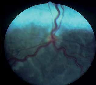
Optic nerve head hypoplasia. The optic nerve head has a reduced diameter and retinal vessels are tortuous. Whitish vermiform lesions are characteristic of concurrent retinal dysplasia.
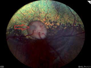
Optic nerve head coloboma, arrow.
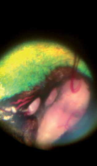
Optic nerve coloboma in a German Shepherd puppy. Retinal vessels change direction as they approach the optic nerve head, appearing to descend into a crater-like depression; the inset marks the vessels.
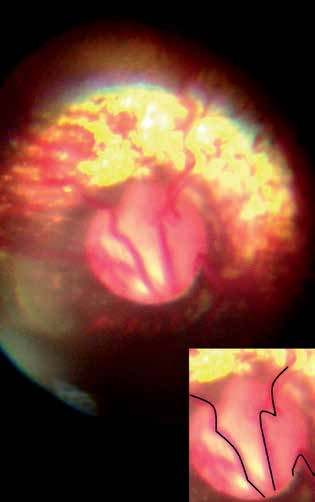
Optic nerve head coloboma.
Adapted from the Atlas of Ocular Diseases in Dogs and Cats by Dr. Siniša D. Grozdanić and colleagues. © Animal Eye Consultants of Iowa. The text and clinical images on this page are original content from the authors' own clinical practice and research, and may not be reproduced, distributed, or repurposed without permission.
Think your pet may have optic nerve hypoplasia/aplasia/coloboma?
Early evaluation gives us the most treatment options and the best chance of preserving vision.