
Optic Nerve Atrophy
Optic nerve atrophy is the end result of neuropathies of many etiologies. It may follow compressive, traumatic, ischemic, inflammatory including infectious or autoimmune, infiltrative, or toxic neuropathy. Optic nerve atrophy may also result from primary disease of other ocular structures, including progressive retinal atrophy, extraocular myositis, and, most commonly according to the source, glaucoma. It can also occur with autoimmune granulomatous meningoencephalitis and meningitis of other causes that produce optic neuritis.
Changes in the optic nerve are characteristic. The optic nerve head becomes smaller because of neuronal loss and develops sharply defined margins. Blood vessels are absent from the disc and its color is pale, light gray, or dark gray. Retinal vessels may be reduced or absent. An eye with optic nerve atrophy is blind and lacks a pupillary light response. Additional ocular or systemic signs depend on the primary cause; for example, glaucoma may also produce increased intraocular pressure, corneal edema, and lens luxation.
Treatment
Diagnosis is established by fundic examination with indirect ophthalmoscopy. Optic nerve atrophy is a consequence or manifestation of another disease, so the primary cause should be identified. This is important to protect the fellow eye when the process is bilateral but asymmetric and to identify potentially life-threatening systemic disease. There is no treatment to restore vision in a blind eye with established optic nerve atrophy; early diagnosis and treatment of the underlying cause are therefore essential.
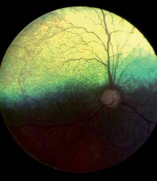
Glaucomatous optic neuropathy, early stage. Optic nerve and retina in a patient with primary hereditary glaucoma. The optic nerve shows mild atrophy with gray coloration and central excavation, but no retinal degeneration or retinal vascular atrophy. Daytime vision is still preserved in this eye.
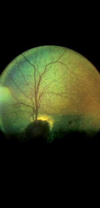
Glaucomatous optic neuropathy, late stage. Optic nerve and retina in a patient with primary hereditary glaucoma. The optic nerve shows marked atrophy and central excavation. Retinal degeneration is not apparent, but mild retinal vascular atrophy is present. This eye is blind.
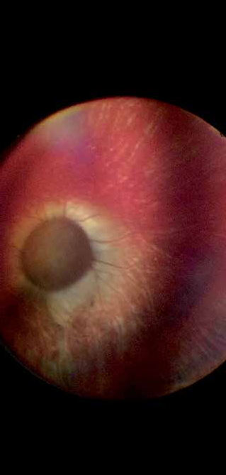
End-stage glaucomatous optic neuropathy. The optic nerve is completely atrophic, as is the choroidal vasculature surrounding it. The optic nerve changes are characteristic of glaucoma, described as optic nerve cupping/capping in the source. Retinal vessels are in an advanced stage of atrophy and are barely visible. This eye is blind.
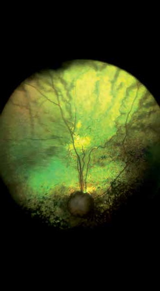
Dog with traumatic head injury after attack by a larger dog. Trauma caused irreversible optic nerve injury and focal retinal injury, commotio retinae.
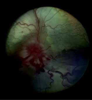
Compressive optic neuropathy caused by an inflammatory process involving the extraocular muscles. The patient became blind. Optic nerve atrophy will develop as the final consequence of ischemia.
Adapted from the Atlas of Ocular Diseases in Dogs and Cats by Dr. Siniša D. Grozdanić and colleagues. © Animal Eye Consultants of Iowa. The text and clinical images on this page are original content from the authors' own clinical practice and research, and may not be reproduced, distributed, or repurposed without permission.
Think your pet may have optic nerve atrophy?
Early evaluation gives us the most treatment options and the best chance of preserving vision.