
Meibomian Gland Adenoma/Epithelioma
A Meibomian gland adenoma is a benign tumor arising from the sebocytes of a Meibomian gland. The tumor develops along the eyelid margin and is usually white to pale pink, less commonly darkly pigmented, and may have smooth margins or a cauliflower-like appearance. Several tumors can occasionally develop on one eyelid. They generally do not cause problems, but a dog may traumatize them by scratching, which can result in secondary bacterial infection. Depending on tumor location, irritation of the cornea and conjunctiva may occur, resulting in keratitis, corneal ulceration, and conjunctivitis.
A Meibomian gland epithelioma is a low-grade malignant tumor rather than a benign tumor and develops from reserve (supporting) cells of the Meibomian gland. Unlike an adenoma, an epithelioma tends to infiltrate surrounding tissues more aggressively, which can make complete surgical excision more difficult. Metastasis is extremely rare. Grossly, the epithelioma closely resembles a Meibomian gland adenoma. It appears along the eyelid margin as a white to pale-pink, occasionally more deeply pigmented growth that may be smooth or cauliflower-like. As with Meibomian gland adenoma, secondary bacterial infection may cause inflammation of the tumor. Depending on its location, corneal and conjunctival irritation may result in keratitis, corneal ulceration, and conjunctivitis. A chalazion may develop as a consequence of mechanical obstruction of Meibomian glands by the tumor.
Meibomian gland adenomas and epitheliomas occur in older dogs and are often seen in Cocker Spaniels, Poodles, Siberian Huskies, Samoyeds, and Shih Tzus. They are extremely rare in cats.
Treatment
Diagnosis is established by histopathologic examination after excision. Tumors are removed by laser or surgical excision.
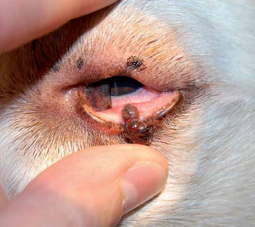
Meibomian gland adenoma of the lower eyelid. Two brown, cauliflower-like tumors are present.
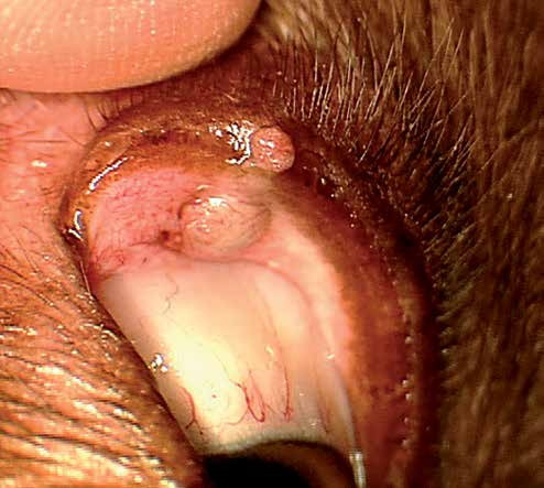
Meibomian gland adenoma of the upper eyelid. A smooth pink mass extends along the length of the Meibomian gland.
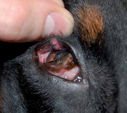
Meibomian gland epithelioma of the upper eyelid.
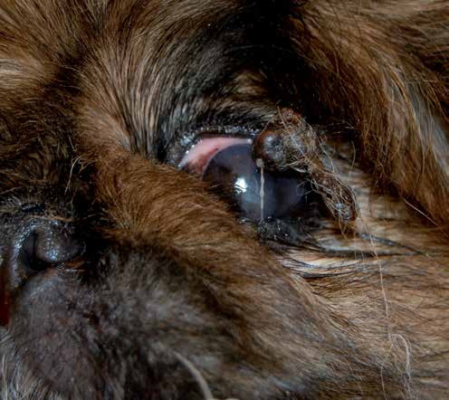
Meibomian gland adenoma of the upper eyelid. Hairs around the tumor are matted with discharge. The tumor caused marked corneal irritation in this dog, and scratching resulted in trauma and infection of the tumor.
Adapted from the Atlas of Ocular Diseases in Dogs and Cats by Dr. Siniša D. Grozdanić and colleagues. © Animal Eye Consultants of Iowa. The text and clinical images on this page are original content from the authors' own clinical practice and research, and may not be reproduced, distributed, or repurposed without permission.
Think your pet may have meibomian gland adenoma/epithelioma?
Early evaluation gives us the most treatment options and the best chance of preserving vision.