
Iris Bombe
Iris bombe is a postinflammatory iris abnormality in which the iris bows or balloons anteriorly. Most commonly after anterior uveitis, accumulated inflammatory cells, fibrin, and fibroblasts cause synechiae between the iris and lens, posterior synechiae, or between iris and cornea, anterior synechiae.
If posterior synechiae form around the entire pupillary margin, the posterior iris adheres to the anterior lens capsule and the central iris bows forward relative to the peripheral iris. The lens may become obscured by the anteriorly displaced iris. Iris bombe may also occur after trauma, glaucoma, lens luxation, or as a complication of cataract surgery, all of which may be associated with uveitis. Iris bombe can cause acute secondary glaucoma by closing the iridocorneal angle and disrupting aqueous-humor flow. Clinically, the anteriorly bowed iris, altered pupil shape, cloudy ocular appearance, and episcleral and conjunctival hyperemia are readily visible.
Treatment
Diagnosis is established by ophthalmic examination. Surgical lysis of iris adhesions, laser iridotomy, and/or lens removal may be attempted, but adhesions tend to recur. Uveitis and glaucoma must be treated appropriately. Prognosis is poor.
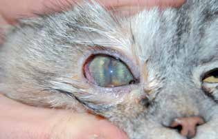
Iris bombe in a cat. Anterior bowing of the iris and an irregular pupil are visible.
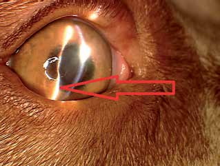
Iris bombe in a dog. During biomicroscopic examination, the slit beam over the iris surface appears wavy rather than straight.
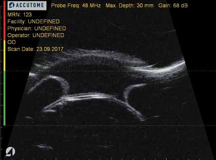
Iris bombe in a dog. Ultrasound image demonstrates an anteriorly bowed iris approaching the cornea.
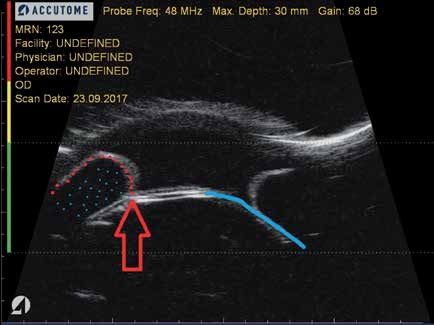
Iris bombe in a dog. Annotated ultrasound image. The red arrow identifies the site of synechia. Impaired aqueous outflow causes fluid to accumulate and displace the iris anteriorly; the blue line represents the normal physiologic iris position.
Adapted from the Atlas of Ocular Diseases in Dogs and Cats by Dr. Siniša D. Grozdanić and colleagues. © Animal Eye Consultants of Iowa. The text and clinical images on this page are original content from the authors' own clinical practice and research, and may not be reproduced, distributed, or repurposed without permission.
Think your pet may have iris bombe?
Early evaluation gives us the most treatment options and the best chance of preserving vision.