
Iridociliary Adenoma/Adenocarcinoma
Iridociliary epithelial tumors, including benign adenoma and malignant adenocarcinoma, are described as the second most common primary intraocular tumors in dogs after melanoma and melanocytoma. They arise from epithelial cells of the iris or ciliary body and occur in middle-aged and older dogs. Clinically they may be segmented or nonsegmented, invasive or noninvasive, solid or cystic, slow-growing masses that are pink or ochre colored. Adenocarcinoma rarely metastasizes but can be markedly invasive. Secondary complications include glaucoma, uveitis, hyphema, and retinal detachment.
Treatment
Definitive diagnosis is made by histopathologic examination of the tumor following enucleation or surgical tumor removal.
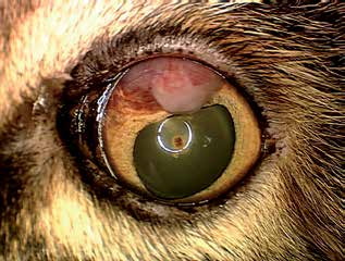
Uveal adenocarcinoma in a cat.
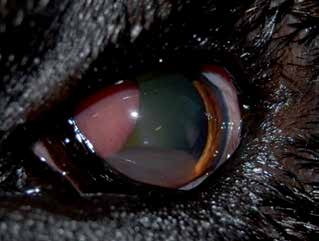
Ciliary body adenoma in a dog.
Adapted from the Atlas of Ocular Diseases in Dogs and Cats by Dr. Siniša D. Grozdanić and colleagues. © Animal Eye Consultants of Iowa. The text and clinical images on this page are original content from the authors' own clinical practice and research, and may not be reproduced, distributed, or repurposed without permission.
Think your pet may have iridociliary adenoma/adenocarcinoma?
Early evaluation gives us the most treatment options and the best chance of preserving vision.