
Internal and External Ophthalmoplegia
Ophthalmoplegia is a neurologic deficit resulting from dysfunction of the oculomotor nerve and/or its nucleus. The oculomotor nerve innervates the dorsal, medial, and ventral rectus muscles, the ventral oblique muscle, and the ipsilateral levator palpebrae superioris. It also contains parasympathetic fibers that innervate intrinsic ocular muscles associated with pupillary function.
Oculomotor nerve lesions may cause internal ophthalmoplegia, characterized by paralysis of the iris and ciliary body with a dilated, nonresponsive pupil; external ophthalmoplegia, characterized by paralysis of extraocular and eyelid muscles with ventrolateral strabismus and ptosis; or complete ophthalmoplegia, in which both internal and external components are affected.
Causes include neoplasia involving the oculomotor nerve, polyneuropathies associated with autoimmune or infectious disease, and idiopathic neuropathies. Ophthalmoplegia may occur as part of cavernous sinus syndrome, in which dysfunction of multiple cranial nerves is present.
Treatment
Testing with dilute pilocarpine can help confirm a deficit in parasympathetic innervation. MRI may help distinguish neoplastic from inflammatory causes, assess the extent of the process, and localize the lesion. Treatment is primarily directed at the underlying cause of ophthalmoplegia.
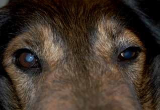
Complete ophthalmoplegia. Ptosis, ventrolateral strabismus, and mydriasis of the left eye are visible.
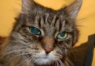
Internal ophthalmoplegia in a cat. Mydriasis of the right pupil is visible.
Adapted from the Atlas of Ocular Diseases in Dogs and Cats by Dr. Siniša D. Grozdanić and colleagues. © Animal Eye Consultants of Iowa. The text and clinical images on this page are original content from the authors' own clinical practice and research, and may not be reproduced, distributed, or repurposed without permission.
Think your pet may have internal and external ophthalmoplegia?
Early evaluation gives us the most treatment options and the best chance of preserving vision.