
Immune-Mediated Retinopathies
The source groups sudden acquired retinal degeneration syndrome (SARDS), immune-mediated retinopathy (IMR), and cancer-associated/paraneoplastic retinopathy (CAR) as immune-mediated retinopathies in which the immune system attacks retinal cells and causes injury.
SARDS is characterized by sudden loss of vision in dogs. The source states that approximately 50% of dogs have accompanying signs such as increased appetite, increased thirst, increased urination, and weight gain, along with altered serum biochemical parameters that resemble hyperadrenocorticism. SARDS most commonly occurs in middle-aged small- to medium-breed dogs. The source describes retinal autoantibodies and an immune-mediated basis. Characteristic findings described are an absent pupillary response to red chromatic light with a preserved response to blue light, an extinguished ERG, and initially minimal or absent visible fundic abnormalities. When abnormalities are present, fundic hyperreflectivity and retinal vascular attenuation may be seen.
Immune-mediated retinopathy is described as similar to SARDS. Approximately 5% of dogs with IMR are stated in the source to have diagnosed cancer. The source proposes that antitumor immune activation may cross-react with similar retinal molecules. When cancer is identified, the condition is termed paraneoplastic retinopathy or cancer-associated retinopathy (CAR). In all three retinopathies, the source describes participation of T lymphocytes, macrophages, B lymphocytes, and autoantibodies.
Treatment
Diagnosis and prognosis are based on history, ophthalmic examination, chromatic pupillary light reflex testing, and ERG. Detailed testing is recommended to determine whether an underlying cancer is present. According to the source, early diagnosis and timely immunosuppressive therapy may allow long-term preservation of vision.
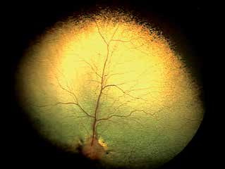
SARDS in a dog. Tapetal hyperreflectivity and retinal vascular attenuation are visible.
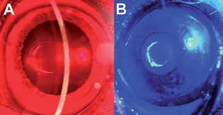
Chromatic pupillary light reflex. The pupil does not respond to red light and remains dilated but constricts in response to blue light.
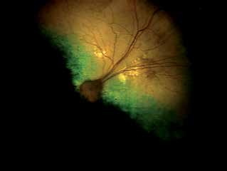
Dog diagnosed with IMR. Localized tapetal hyperreflective areas and attenuation of retinal vessels are visible.
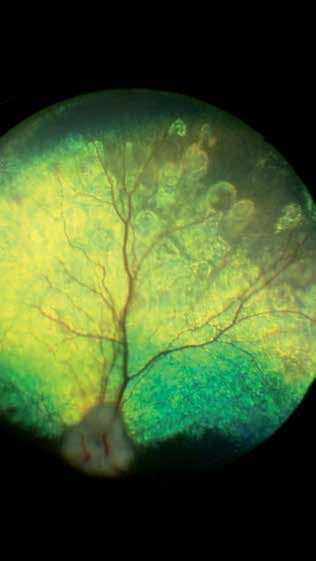
Focal degenerative changes are present in the perivascular region in this dog with autoimmune retinopathy, specifically paraneoplastic/cancer-associated retinopathy, described in the source as most likely associated with mammary adenocarcinoma.
Related in-depth guide
SARDS / IMR
Our full guide, including treatment and surgical options
Adapted from the Atlas of Ocular Diseases in Dogs and Cats by Dr. Siniša D. Grozdanić and colleagues. © Animal Eye Consultants of Iowa. The text and clinical images on this page are original content from the authors' own clinical practice and research, and may not be reproduced, distributed, or repurposed without permission.
Think your pet may have immune-mediated retinopathies?
Early evaluation gives us the most treatment options and the best chance of preserving vision.