
Hypertensive Retinopathy
Hypertension, defined in the source as persistent systolic blood pressure above 160 mmHg, can cause numerous ocular lesions. Dogs and cats may develop primary hypertension without another systemic disorder, but hypertension is often secondary to kidney disease, endocrinopathies such as hyperadrenocorticism, diabetes mellitus, and hyperthyroidism, or neoplasia such as pheochromocytoma and lymphoma.
Hypertension causes arterial vasoconstriction followed by vascular occlusion and necrosis and ultimately increased vascular permeability. The source states that more than 60% of hypertensive patients have ophthalmic consequences and recommends regular ophthalmic examinations. Common complications include subretinal edema, retinal hemorrhage, retinal detachment, vitreous hemorrhage, hyphema, glaucoma, optic disc edema, and eventually optic nerve atrophy.
Patients commonly present with sudden loss of vision, which may be reversible if systemic hypertension is controlled. In cats, detached retina may return toward its normal position after blood pressure is stabilized. The source describes systemic hypertension as the most common cause of acute blindness in cats and recommends blood-pressure measurement in every patient with sudden vision loss.
Treatment
When ophthalmic lesions suggest hypertension, blood pressure should be measured after the patient has acclimated and calmed. Diagnostic investigation should identify and address the cause of hypertension. Antihypertensive therapy includes calcium-channel blockers and ACE inhibitors as described in the source. Ocular complications such as hyphema, retinal detachment, and glaucoma should be treated appropriately.
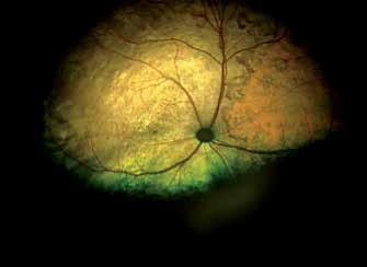
Hypertensive retinopathy in a cat with hyperthyroidism. The cat is blind. Optic nerve head atrophy, small subretinal hemorrhages, and a large orange area representing accumulated fluid between choroid and retina are visible.
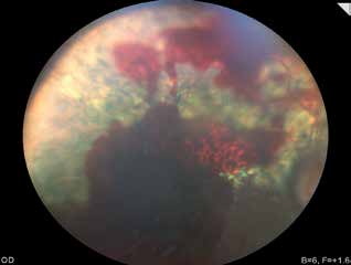
Hypertensive retinopathy in a dog. Retinal hemorrhages developed secondary to hypertension caused by pheochromocytoma.
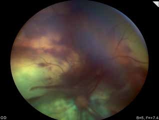
Hypertensive retinopathy in a cat. Subretinal hemorrhages and retinal folds caused by focal retinal detachment are present.
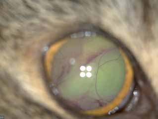
Hypertensive retinopathy in a cat. Retinal vessels are visible through the dilated pupil, indicating retinal detachment.
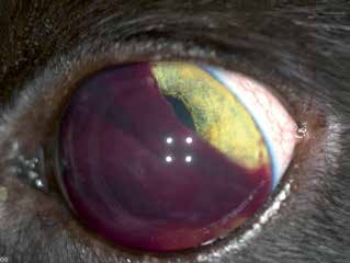
Hypertensive retinopathy in a cat. Retinal detachment resulted in intraocular hemorrhage; blood is also present in the anterior chamber as hyphema.
Related in-depth guide
Retinal Disorders
Our full guide, including treatment and surgical options
Adapted from the Atlas of Ocular Diseases in Dogs and Cats by Dr. Siniša D. Grozdanić and colleagues. © Animal Eye Consultants of Iowa. The text and clinical images on this page are original content from the authors' own clinical practice and research, and may not be reproduced, distributed, or repurposed without permission.
Think your pet may have hypertensive retinopathy?
Early evaluation gives us the most treatment options and the best chance of preserving vision.