
Horner Syndrome
Horner syndrome is a group of clinical signs caused by dysfunction of sympathetic innervation to the eye. Sympathetic pathways innervate ocular and periocular structures involved in pupil dilation, orbital smooth-muscle tone, and eyelid position; in cats they also contribute to smooth-muscle control of the third eyelid.
Partial or complete interruption of sympathetic innervation may result from trauma to the neck or thorax, including blunt trauma and bites; neoplasia; intervertebral disease; inflammatory disease; middle- or inner-ear disease; polyps, particularly in cats; or metabolic disease such as diabetes mellitus. The source describes allergic ear disease as the most common cause of Horner syndrome in dogs. Horner syndrome can also follow certain surgical procedures, while in many patients no cause is identified and the syndrome is classified as idiopathic.
Signs develop suddenly and include ptosis, protrusion of the third eyelid, miosis, enophthalmos, and conjunctival hyperemia. Signs are more commonly unilateral but can be bilateral and may occur in dogs of any age or breed. The source notes reports of somewhat greater frequency in Labrador Retrievers, Collies, Shetland Sheepdogs, and Doberman Pinschers.
Treatment
Topical 10% phenylephrine is used to help differentiate Horner syndrome from other causes of miosis. Phenylephrine testing may assist lesion localization to central, preganglionic, or postganglionic sympathetic pathways. According to the source, a positive response supports postganglionic Horner syndrome, which is described as the most commonly diagnosed localization. Further investigation should seek the underlying cause and may include ear and eye examination, skull and thoracic radiography, MRI, and CT. Some cases resolve spontaneously over weeks to months. Prognosis and treatment depend on the primary cause. Topical sympathomimetic therapy may be used when the condition produces functional visual difficulty.
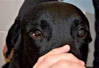
Horner syndrome in a dog. In the right eye of this Labrador Retriever, enophthalmos, miosis, and third eyelid protrusion are visible.
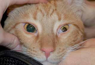
Horner syndrome in a cat. The left eye shows enophthalmos, third eyelid protrusion, ptosis, and miosis.
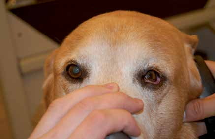
Horner syndrome in a dog. The left eye shows ptosis, enophthalmos, third eyelid protrusion, miosis, and lower eyelid ectropion.
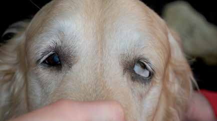
Horner syndrome in a dog. The left eye shows ptosis, enophthalmos, third eyelid protrusion, and lower eyelid ectropion.
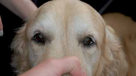
The same dog as in the previous photograph, 15 minutes after phenylephrine eye drops.
Adapted from the Atlas of Ocular Diseases in Dogs and Cats by Dr. Siniša D. Grozdanić and colleagues. © Animal Eye Consultants of Iowa. The text and clinical images on this page are original content from the authors' own clinical practice and research, and may not be reproduced, distributed, or repurposed without permission.
Think your pet may have horner syndrome?
Early evaluation gives us the most treatment options and the best chance of preserving vision.