
Eyelid Injuries
The eyelids have an external component, composed of skin and the orbicularis oculi muscle, and an internal component, composed of conjunctiva and connective tissue. Mechanical injuries to the eyelids most commonly occur as lacerations. In both dogs and cats, they are often seen after fights, hunting, or outdoor play. Lacerations may be superficial, involving only the eyelid skin, or may involve all layers of the eyelid.
An eyelid laceration should be taken seriously regardless of how harmless it may initially appear. Because of contraction of the orbicularis oculi muscle, a small laceration may develop into a large tissue defect. Likewise, newly formed scar tissue may cause inward or outward rolling of the eyelid. Any anatomical abnormality of the eyelid disrupts its function and may consequently damage the cornea. Such damage is most often caused by hairs originating from eyelid skin that, because of tissue injury, come into contact with the ocular surface, or by scar tissue formed along the eyelid margin that irritates sensitive ocular structures.
Treatment
Whenever an eyelid laceration is present, a thorough ophthalmic examination should be performed to determine whether there are hidden injuries to internal ocular structures or a foreign body within the eye. The wound should be surgically treated as soon as possible and the eyelid margin restored to its pre-injury anatomical position. The wound should not be allowed to heal spontaneously because an anatomical defect may form that compromises the physiological role of the eyelids, and because bacterial infection may occur. For these reasons, eyelid lacerations should be surgically repaired as soon as possible. If needed, an Elizabethan collar is placed after the procedure to prevent the dog from scratching and disrupting the sutures.
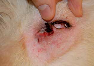
Lower eyelid laceration sustained by this dog after a fight with another dog. One third of the lower eyelid margin, 2-3 mm in width, is detached and requires urgent reconstruction.
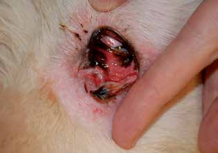
Eyelid laceration. The same dog after detailed examination, demonstrating the true severity of the laceration.
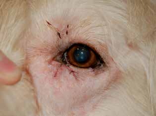
Eyelid laceration. The same dog after surgery. Repositioning was performed correctly and the eyelid margin was restored to the proper anatomical position, which is necessary for normal eyelid function.
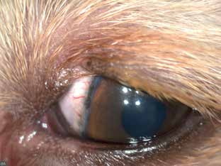
Dog photographed after an upper eyelid laceration that healed spontaneously. Note the contact between hairs of the upper eyelid and the cornea caused by the defect in the eyelid margin.
Adapted from the Atlas of Ocular Diseases in Dogs and Cats by Dr. Siniša D. Grozdanić and colleagues. © Animal Eye Consultants of Iowa. The text and clinical images on this page are original content from the authors' own clinical practice and research, and may not be reproduced, distributed, or repurposed without permission.
Think your pet may have eyelid injuries?
Early evaluation gives us the most treatment options and the best chance of preserving vision.