
Eyelid Inflammation (Blepharitis)
Blepharitis may be focal, as in hordeolum and granulomatous blepharitis, with inflammation of one or more Meibomian glands, or it may develop as a diffuse disorder causing inflammation of the entire eyelid margin. It may occur as a primary problem or as one manifestation of generalized skin disease. Accordingly, the causes of blepharitis are the same as those of dermatitis.
Infectious causes include parasites (Demodex, Sarcoptes, Cuterebra larvae, Hypoderma larvae), bacteria (Staphylococcus spp., Streptococcus spp., and others), fungi (Microsporum canis, Microsporum gypseum, Trichophyton mentagrophytes, and Malassezia pachydermatis), and certain protozoa such as Leishmania infantum. Causes are also frequently immunologic, ranging from allergies to autoimmune disease, as well as vascular and nutritional. Individual causes of blepharitis are described separately in subsequent sections.
Clinical signs vary according to the cause and duration of disease. The most common findings include marked eyelid swelling and erythema, blepharospasm, and tearing. Blepharitis is frequently accompanied by conjunctival inflammation (blepharoconjunctivitis). Periocular alopecia, erosions, scales, crusts, exudation, and scarring may also be present.
Treatment
For treatment to be successful, the exact cause must be identified. If bacterial or fungal blepharitis is suspected, cytologic and microbiologic examinations are performed. Demodicosis is diagnosed by skin scraping, and leishmaniasis by serology or PCR testing. Atopic dermatitis is confirmed by intradermal skin testing, and autoimmune diseases by histopathologic examination of a biopsy specimen. Each cause requires specific medical therapy, which may be local and/or systemic. Duration depends on the cause and may extend for months or even lifelong in autoimmune disease.
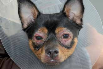
Blepharitis in a dog. Alopecia, edema, hypopigmentation, erosions, and crusts are present.
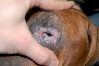
Blepharitis in a dog. Alopecia, edema, hypopigmentation, and erosions are visible.
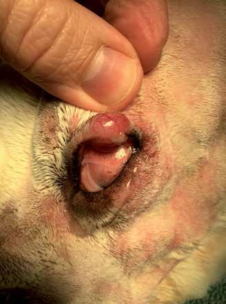
Focal granulomatous blepharitis that destroyed the central portion of the upper eyelid and caused corneal irritation.
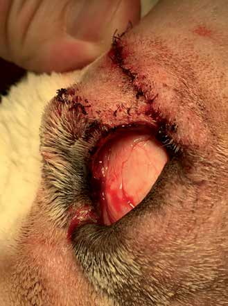
Focal granulomatous blepharitis after surgical resection and reconstruction (H-plasty). The dog from the previous figure shortly after surgery.
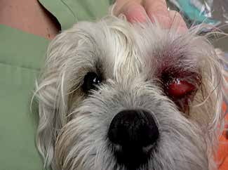
Bacterial blepharitis/cellulitis of the left upper eyelid in a dog. Two weeks of systemic antibiotic and nonsteroidal anti-inflammatory therapy resulted in resolution.
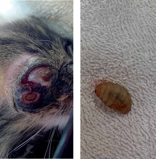
Blepharitis with eyelid damage and corneal infection in a cat as a result of infestation with insect larvae.
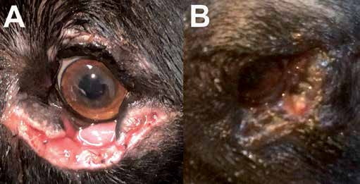
A) Necrotizing fasciitis that developed after entropion surgery. Necrosis of the skin and subcutaneous tissue and dehiscence of the surgical wound were accompanied by dramatic hematologic changes (marked neutrophilia, leukocytosis, anemia, and thrombocytopenia). B) Surgical wound treatment and closure of the defect led to normalization of neutrophil and platelet counts. Partial wound dehiscence was detected on the seventh day after surgery, most likely as a result of persistent necrosis of the subcutaneous tissue. This is one of the rare eyelid conditions in which a fatal outcome is possible if aggressive antibiotic therapy and surgical wound management are delayed.
Adapted from the Atlas of Ocular Diseases in Dogs and Cats by Dr. Siniša D. Grozdanić and colleagues. © Animal Eye Consultants of Iowa. The text and clinical images on this page are original content from the authors' own clinical practice and research, and may not be reproduced, distributed, or repurposed without permission.
Think your pet may have eyelid inflammation (blepharitis)?
Early evaluation gives us the most treatment options and the best chance of preserving vision.